8CTJ | pdb_00008ctj

Cryo-EM structure of TMEM87A


Experimental Data Snapshot

  • Method: ELECTRON MICROSCOPY
  • Resolution: 4.74 Å
  • Aggregation State: PARTICLE 
  • Reconstruction Method: SINGLE PARTICLE 

wwPDB Validation 3D Report Full Report

Validation slider image for 8CTJ

This is version 1.1 of the entry. See complete history

Literature

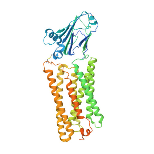
Structure of the GOLD-domain seven-transmembrane helix protein family member TMEM87A.

Hoel, C.M.Zhang, L.Brohawn, S.G.

(2022) Elife 11

  • DOI: https://doi.org/10.7554/eLife.81704
  • Primary Citation Related Structures: 
    8CTJ

  • PubMed Abstract: 

    TMEM87s are eukaryotic transmembrane proteins with two members (TMEM87A and TMEM87B) in humans. TMEM87s have proposed roles in protein transport to and from the Golgi, as mechanosensitive ion channels, and in developmental signaling. TMEM87 disruption has been implicated in cancers and developmental disorders. To better understand TMEM87 structure and function, we determined a cryo-EM structure of human TMEM87A in lipid nanodiscs. TMEM87A consists of a Golgi-dynamics (GOLD) domain atop a membrane-spanning seven-transmembrane helix domain with a large cavity open to solution and the membrane outer leaflet. Structural and functional analyses suggest TMEM87A may not function as an ion channel or G-protein coupled receptor. We find TMEM87A shares its characteristic domain arrangement with seven other proteins in humans; three that had been identified as evolutionary related (TMEM87B, GPR107, and GPR108) and four previously unrecognized homologs (GPR180, TMEM145, TMEM181, and WLS). Among these structurally related GO LD domain s even- t ransmembrane helix (GOST) proteins, WLS is best characterized as a membrane trafficking and secretion chaperone for lipidated Wnt signaling proteins. We find key structural determinants for WLS function are conserved in TMEM87A. We propose TMEM87A and structurally homologous GOST proteins could serve a common role in trafficking membrane-associated cargo.


  • Organizational Affiliation
    • Department of Molecular and Cell Biology, University of California, Berkeley, Berkeley, United States.

Macromolecule Content 

  • Total Structure Weight: 65.26 kDa 
  • Atom Count: 3,312 
  • Modeled Residue Count: 401 
  • Deposited Residue Count: 564 
  • Unique protein chains: 1

Macromolecules

Find similar proteins by:|  3D Structure
Entity ID: 1
MoleculeChains  Sequence LengthOrganismDetailsImage
Transmembrane protein 87A564Homo sapiensMutation(s): 0 
Gene Names: TMEM87APSEC0094
Membrane Entity: Yes 
UniProt & NIH Common Fund Data Resources
Find proteins for Q8NBN3 (Homo sapiens)
Explore Q8NBN3 
Go to UniProtKB:  Q8NBN3
PHAROS:  Q8NBN3
GTEx:  ENSG00000103978 
Entity Groups
Sequence Clusters30% Identity50% Identity70% Identity90% Identity95% Identity100% Identity
UniProt GroupQ8NBN3
Sequence Annotations
Expand
Reference Sequence

Small Molecules

Ligands 1 Unique
IDChains Name / Formula / InChI Key2D Diagram3D Interactions
PEE

Query on PEE


Download:Ideal Coordinates CCD File
B [auth A]1,2-dioleoyl-sn-glycero-3-phosphoethanolamine
C41 H78 N O8 P
MWRBNPKJOOWZPW-NYVOMTAGSA-N

Experimental Data & Validation

Experimental Data

  • Method: ELECTRON MICROSCOPY
  • Resolution: 4.74 Å
  • Aggregation State: PARTICLE 
  • Reconstruction Method: SINGLE PARTICLE 

Structure Validation

View Full Validation Report


Entry History 

& Funding Information

Deposition Data


Funding OrganizationLocationGrant Number
National Institutes of Health/National Institute of General Medical Sciences (NIH/NIGMS)United StatesDP2GM123496

Revision History  (Full details and data files)

  • Version 1.0: 2022-07-20
    Type: Initial release
  • Version 1.1: 2022-11-30
    Changes: Database references