Novel exported fusion enzymes with chorismate mutase and cyclohexadienyl dehydratase activity: Shikimate pathway enzymes teamed up in no man's land.
Stocker, C., Khatanbaatar, T., Bressan, L., Wurth-Roderer, K., Cordara, G., Krengel, U., Kast, P.(2023) J Biological Chem 299: 105161-105161
- PubMed: 37586588 Search on PubMedSearch on PubMed Central
- DOI: https://doi.org/10.1016/j.jbc.2023.105161
- Primary Citation Related Structures:
8CQ3, 8CQ4, 8CQ6 - PubMed Abstract:
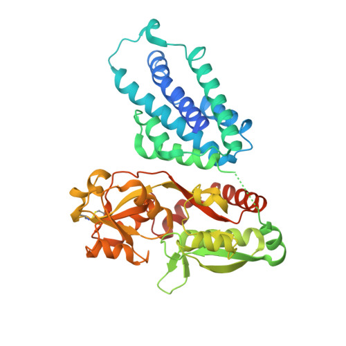
Chorismate mutase (CM) and cyclohexadienyl dehydratase (CDT) catalyze two subsequent reactions in the intracellular biosynthesis of l-phenylalanine (Phe). Here, we report the discovery of novel and extremely rare bifunctional fusion enzymes, consisting of fused CM and CDT domains, which are exported from the cytoplasm. Such enzymes were found in only nine bacterial species belonging to non-pathogenic γ- or β-Proteobacteria. In γ-proteobacterial fusion enzymes, the CM domain is N-terminal to the CDT domain, whereas the order is inverted in β-Proteobacteria. The CM domains share 15% to 20% sequence identity with the AroQ γ class CM holotype of Mycobacterium tuberculosis (∗MtCM), and the CDT domains 40% to 60% identity with the exported monofunctional enzyme of Pseudomonas aeruginosa (PheC). In vitro kinetics revealed a K m <7 μM, much lower than for ∗MtCM, whereas kinetic parameters are similar for CDT domains and PheC. There is no feedback inhibition of CM or CDT by the pathway's end product Phe, and no catalytic benefit of the domain fusion compared with engineered single-domain constructs. The fusion enzymes of Aequoribacter fuscus, Janthinobacterium sp. HH01, and Duganella sacchari were crystallized and their structures refined to 1.6, 1.7, and 2.4 Å resolution, respectively. Neither the crystal structures nor the size-exclusion chromatography show evidence for substrate channeling or higher oligomeric structure that could account for the cooperation of CM and CDT active sites. The genetic neighborhood with genes encoding transporter and substrate binding proteins suggests that these exported bifunctional fusion enzymes may participate in signaling systems rather than in the biosynthesis of Phe.
- Laboratory of Organic Chemistry, D-CHAB, ETH Zurich, Zurich, Switzerland.
Organizational Affiliation: