Actin arginylation alters myosin engagement and F-actin patterning despite structural conservation.
Pinto, C.S., Bakker, S.E., Suchenko, A., Kolodny, I.M., Hussain, H., Hatano, T., Sampath, K., Chinthalapudi, K., Heissler, S.M., Mishima, M., Balasubramanian, M.(2026) J Cell Biol 225
- PubMed: 41236477 Search on PubMedSearch on PubMed Central
- DOI: https://doi.org/10.1083/jcb.202409067
- Primary Citation Related Structures:
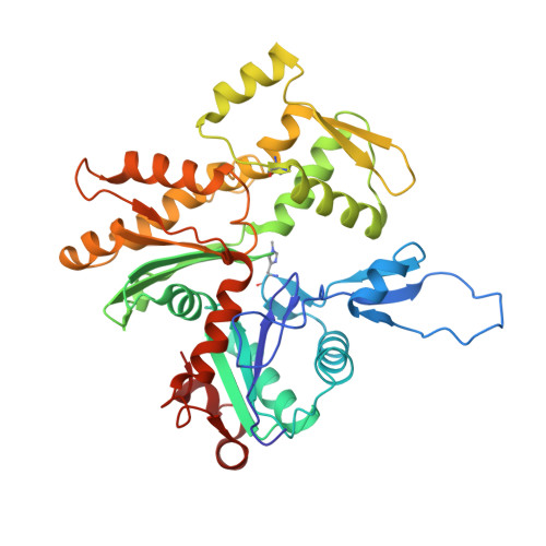
8COG - PubMed Abstract:
Actin is a conserved protein with crucial roles in cell polarity, division, and muscle contraction. Its function is regulated in part by posttranslational modifications, one of which is N-terminal arginylation. What is the structure of arginylated-β-actin (R-β-actin), and how does it regulate F-actin function? Here we report the 3.6 Å structures of ADP-R-β-actin filaments, which are nearly identical to that of non-arginylated F-actin. In vitro assays reveal that the interaction between myosin-II and actin is altered upon actin arginylation, characterized by frequent detachment of R-actin filaments from myosin-II. In vivo, replacement of the only actin gene in Schizosaccharomyces pombe with a synthetic gene encoding R-Sp-actin reduces Arp2/3-based actin patches while thickening formin-induced actin cables. Consistent with defective interactions between myosin-II and R-actin filaments, assembly and constriction of the cytokinetic actomyosin ring are perturbed in R-Sp-actin cells. Thus, despite structural similarity of arginylated and non-arginylated actin filaments, actin arginylation affects F-actin assortment into distinct subcellular structures and its interaction with myosin-II.
- Centre for Mechanochemical Cell Biology and Division of Biomedical Sciences, Warwick Medical School, Coventry, UK.
Organizational Affiliation: