8CH1

PBP AccA from A. vitis S4 in complex with Agrocinopine A


Experimental Data Snapshot

  • Method: X-RAY DIFFRACTION
  • Resolution: 1.50 Å
  • R-Value Free: 0.202 
  • R-Value Work: 0.174 
  • R-Value Observed: 0.175 

wwPDB Validation   3D Report Full Report


Ligand Structure Quality Assessment 


This is version 1.1 of the entry. See complete history


Literature

A highly conserved ligand-binding site for AccA transporters of antibiotic and quorum-sensing regulator in Agrobacterium leads to a different specificity.

Morera, S.Vigouroux, A.Aumont-Nicaise, M.Ahmar, M.Meyer, T.El Sahili, A.Deicsics, G.Gonzalez-Mula, A.Li, S.Dore, J.Sirigu, S.Legrand, P.Penot, C.Andre, F.Faure, D.Soulere, L.Queneau, Y.Vial, L.

(2024) Biochem J 481: 93-117

  • DOI: https://doi.org/10.1042/BCJ20230273
  • Primary Citation of Related Structures:  
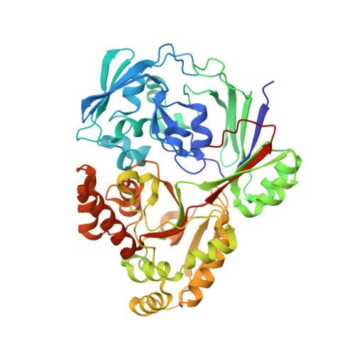
    8C6R, 8C6U, 8C6W, 8C6Y, 8C75, 8CAW, 8CAY, 8CB9, 8CDO, 8CH1, 8CH2, 8CH3, 8CHC, 8CI6, 8CJU, 8CKD, 8CKE, 8CKO

  • PubMed Abstract: 

    Plants genetically modified by the pathogenic Agrobacterium strain C58 synthesize agrocinopines A and B, whereas those modified by the pathogenic strain Bo542 produce agrocinopines C and D. The four agrocinopines (A, B, C and D) serve as nutrients by agrobacteria and signaling molecule for the dissemination of virulence genes. They share the uncommon pyranose-2-phosphate motif, represented by the l-arabinopyranose moiety in agrocinopines A/B and the d-glucopyranose moiety in agrocinopines C/D, also found in the antibiotic agrocin 84. They are imported into agrobacterial cytoplasm via the Acc transport system, including the solute-binding protein AccA coupled to an ABC transporter. We have previously shown that unexpectedly, AccA from strain C58 (AccAC58) recognizes the pyranose-2-phosphate motif present in all four agrocinopines and agrocin 84, meaning that strain C58 is able to import agrocinopines C/D, originating from the competitor strain Bo542. Here, using agrocinopine derivatives and combining crystallography, affinity and stability measurements, modeling, molecular dynamics, in vitro and vivo assays, we show that AccABo542 and AccAC58 behave differently despite 75% sequence identity and a nearly identical ligand binding site. Indeed, strain Bo542 imports only compounds containing the d-glucopyranose-2-phosphate moiety, and with a lower affinity compared with strain C58. This difference in import efficiency makes C58 more competitive than Bo542 in culture media. We can now explain why Agrobacterium/Allorhizobium vitis strain S4 is insensitive to agrocin 84, although its genome contains a conserved Acc transport system. Overall, our work highlights AccA proteins as a case study, for which stability and dynamics drive specificity.


  • Organizational Affiliation

    Université Paris-Saclay, CEA, CNRS, Institute for Integrative Biology of the Cell (I2BC), 91198 Gif-sur-Yvette, France.


Macromolecules
Find similar proteins by:  (by identity cutoff)  |  3D Structure
Entity ID: 1
MoleculeChains Sequence LengthOrganismDetailsImage
Agrocinopine utilization periplasmic binding protein AccA499Allorhizobium ampelinum S4Mutation(s): 0 
UniProt
Find proteins for B9JXG4 (Allorhizobium ampelinum (strain ATCC BAA-846 / DSM 112012 / S4))
Explore B9JXG4 
Go to UniProtKB:  B9JXG4
Entity Groups  
Sequence Clusters30% Identity50% Identity70% Identity90% Identity95% Identity100% Identity
UniProt GroupB9JXG4
Sequence Annotations
Expand
  • Reference Sequence
Oligosaccharides

Help

Entity ID: 2
MoleculeChains Length2D Diagram Glycosylation3D Interactions
beta-D-fructofuranose-(2-1)-alpha-D-glucopyranose
B, C
2N/A
Glycosylation Resources
GlyTouCan:  G05551OP
GlyCosmos:  G05551OP
Small Molecules
Ligands 4 Unique
IDChains Name / Formula / InChI Key2D Diagram3D Interactions
VDF (Subject of Investigation/LOI)
Query on VDF

Download Ideal Coordinates CCD File 
D [auth A]2-O-phosphono-beta-L-arabinopyranose
C5 H11 O8 P
QIAIUTJEZJIPSH-KLVWXMOXSA-N
LAO (Subject of Investigation/LOI)
Query on LAO

Download Ideal Coordinates CCD File 
E [auth A]2-O-phosphono-alpha-L-arabinopyranose
C5 H11 O8 P
QIAIUTJEZJIPSH-QMKXCQHVSA-N
PEG
Query on PEG

Download Ideal Coordinates CCD File 
I [auth A]DI(HYDROXYETHYL)ETHER
C4 H10 O3
MTHSVFCYNBDYFN-UHFFFAOYSA-N
EDO
Query on EDO

Download Ideal Coordinates CCD File 
F [auth A],
G [auth A],
H [auth A]
1,2-ETHANEDIOL
C2 H6 O2
LYCAIKOWRPUZTN-UHFFFAOYSA-N
Biologically Interesting Molecules (External Reference) 1 Unique
Experimental Data & Validation

Experimental Data

  • Method: X-RAY DIFFRACTION
  • Resolution: 1.50 Å
  • R-Value Free: 0.202 
  • R-Value Work: 0.174 
  • R-Value Observed: 0.175 
  • Space Group: P 21 21 21
Unit Cell:
Length ( Å )Angle ( ˚ )
a = 47.1α = 90
b = 60.61β = 90
c = 151.28γ = 90
Software Package:
Software NamePurpose
BUSTERrefinement
XDSdata reduction
XSCALEdata scaling
PHASERphasing

Structure Validation

View Full Validation Report


Ligand Structure Quality Assessment 


Entry History & Funding Information

Deposition Data


Funding OrganizationLocationGrant Number
Not funded--

Revision History  (Full details and data files)

  • Version 1.0: 2024-01-24
    Type: Initial release
  • Version 1.1: 2024-02-07
    Changes: Database references