The homodimerization domain of the Stl repressor is crucial for efficient inhibition of mycobacterial dUTPase.
Toth, Z.S., Leveles, I., Nyiri, K., Nagy, G.N., Harmat, V., Jaroentomeechai, T., Ozohanics, O., Miller, R.L., Alvarez, M.B., Vertessy, B.G., Benedek, A.(2024) Sci Rep 14: 27171-27171
- PubMed: 39511242 Search on PubMedSearch on PubMed Central
- DOI: https://doi.org/10.1038/s41598-024-76349-2
- Primary Citation Related Structures:
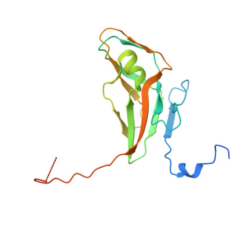
8CGA, 8P8O - PubMed Abstract:
The dUTPase is a key DNA repair enzyme in Mycobacterium tuberculosis, and it may serve as a novel promising anti-tuberculosis target. Stl repressor from Staphylococcus aureus was shown to bind to and inhibit dUTPases from various sources, and its expression in mycobacterial cells interfered with cell growth. To fine-tune and optimize Stl-induced inhibition of mycobacterial dUTPase, we aimed to decipher the molecular details of this interaction. Structural background of the complex between dUTPase and a truncated Stl lacking the repressor C-terminal homodimerization domain has been described, however, the effects of this truncation of Stl on enzyme binding and inhibition are still not known. Using several independent biophysical, structural and enzyme kinetic methods, here we show that lack of the repressor homodimerization domain strongly perturbs both enzyme binding and inhibition. We also investigated the role of a mycobacteria-specific loop in the Stl-interaction. Our results show that removal of this loop leads to a ten-fold increase in the apparent inhibition constant of Stl. We present a high-resolution three-dimensional structure of mycobacterial dUTPase lacking the genus-specific loop for structural insight. Our present data suggest that potent inhibition of mycobacterial dUTPase by Stl requires the wild-type full-length protein context.
- Institute of Molecular Life Sciences, HUN-REN Research Centre for Natural Sciences, Budapest, Hungary. toth.zoe@ttk.hu.
Organizational Affiliation: