Cell-active small molecule inhibitors validate the SNM1A DNA repair nuclease as a cancer target.
Bielinski, M., Henderson, L.R., Yosaatmadja, Y., Swift, L.P., Baddock, H.T., Bowen, M.J., Brem, J., Jones, P.S., McElroy, S.P., Morrison, A., Speake, M., van Boeckel, S., van Doornmalen, E., van Groningen, J., van den Hurk, H., Gileadi, O., Newman, J.A., McHugh, P.J., Schofield, C.J.(2024) Chem Sci 15: 8227-8241
- PubMed: 38817593 Search on PubMedSearch on PubMed Central
- DOI: https://doi.org/10.1039/d4sc00367e
- Primary Citation Related Structures:
8C8B, 8C8D, 8C8S, 8CEW, 8CF0, 8CG9 - PubMed Abstract:
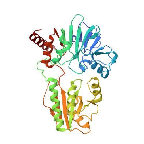
The three human SNM1 metallo-β-lactamase fold nucleases (SNM1A-C) play key roles in DNA damage repair and in maintaining telomere integrity. Genetic studies indicate that they are attractive targets for cancer treatment and to potentiate chemo- and radiation-therapy. A high-throughput screen for SNM1A inhibitors identified diverse pharmacophores, some of which were shown by crystallography to coordinate to the di-metal ion centre at the SNM1A active site. Structure and turnover assay-guided optimization enabled the identification of potent quinazoline-hydroxamic acid containing inhibitors, which bind in a manner where the hydroxamic acid displaces the hydrolytic water and the quinazoline ring occupies a substrate nucleobase binding site. Cellular assays reveal that SNM1A inhibitors cause sensitisation to, and defects in the resolution of, cisplatin-induced DNA damage, validating the tractability of MBL fold nucleases as cancer drug targets.
- Chemistry Research Laboratory, Department of Chemistry and the Ineos Oxford Institute for Antimicrobial Research, University of Oxford Mansfield Road Oxford OX1 3TA UK christopher.schofield@chem.ox.ac.uk.
Organizational Affiliation: