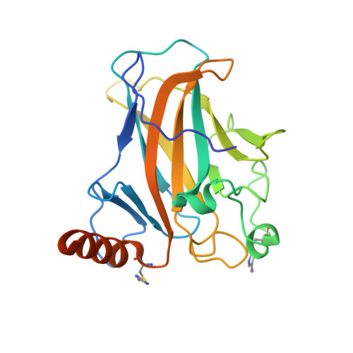
8CG7

Structure of p53 cancer mutant Y220C with arylation at Cys182 and Cys277


Experimental Data Snapshot

  • Method: X-RAY DIFFRACTION
  • Resolution: 1.53 Å
  • R-Value Free: 0.187 
  • R-Value Work: 0.158 
  • R-Value Observed: 0.159 

Starting Model: experimental
View more details

wwPDB Validation   3D Report Full Report


Ligand Structure Quality Assessment 


This is version 2.0 of the entry. See complete history


Literature

Structure-Reactivity Studies of 2-Sulfonylpyrimidines Allow Selective Protein Arylation.

Pichon, M.M.Drelinkiewicz, D.Lozano, D.Moraru, R.Hayward, L.J.Jones, M.McCoy, M.A.Allstrum-Graves, S.Balourdas, D.I.Joerger, A.C.Whitby, R.J.Goldup, S.M.Wells, N.Langley, G.J.Herniman, J.M.Baud, M.G.J.

(2023) Bioconjug Chem 34: 1679-1687

  • DOI: https://doi.org/10.1021/acs.bioconjchem.3c00322
  • Primary Citation of Related Structures:  
    8CG7

  • PubMed Abstract: 

    Protein arylation has attracted much attention for developing new classes of bioconjugates with improved properties. Here, we have evaluated 2-sulfonylpyrimidines as covalent warheads for the mild, chemoselective, and metal free cysteine S -arylation. 2-Sulfonylpyrimidines react rapidly with cysteine, resulting in stable S -heteroarylated adducts at neutral pH. Fine tuning the heterocyclic core and exocyclic leaving group allowed predictable S N Ar reactivity in vitro , covering >9 orders of magnitude. Finally, we achieved fast chemo- and regiospecific arylation of a mutant p53 protein and confirmed arylation sites by protein X-ray crystallography. Hence, we report the first example of a protein site specifically S -arylated with iodo-aromatic motifs. Overall, this study provides the most comprehensive structure-reactivity relationship to date on heteroaryl sulfones and highlights 2-sulfonylpyrimidine as a synthetically tractable and protein compatible covalent motif for targeting reactive cysteines, expanding the arsenal of tunable warheads for modern covalent ligand discovery.


  • Organizational Affiliation

    School of Chemistry, University of Southampton, Highfield, SO17 1BJ Southampton, United Kingdom.


Macromolecules
Find similar proteins by:  (by identity cutoff)  |  3D Structure
Entity ID: 1
MoleculeChains Sequence LengthOrganismDetailsImage
Cellular tumor antigen p53
A, B
219Homo sapiensMutation(s): 5 
Gene Names: TP53P53
UniProt & NIH Common Fund Data Resources
Find proteins for P04637 (Homo sapiens)
Explore P04637 
Go to UniProtKB:  P04637
PHAROS:  P04637
GTEx:  ENSG00000141510 
Entity Groups  
Sequence Clusters30% Identity50% Identity70% Identity90% Identity95% Identity100% Identity
UniProt GroupP04637
Sequence Annotations
Expand
  • Reference Sequence
Experimental Data & Validation

Experimental Data

  • Method: X-RAY DIFFRACTION
  • Resolution: 1.53 Å
  • R-Value Free: 0.187 
  • R-Value Work: 0.158 
  • R-Value Observed: 0.159 
  • Space Group: P 21 21 21
Unit Cell:
Length ( Å )Angle ( ˚ )
a = 64.881α = 90
b = 71.046β = 90
c = 104.887γ = 90
Software Package:
Software NamePurpose
PHENIXrefinement
XDSdata reduction
Aimlessdata scaling
PHENIXphasing

Structure Validation

View Full Validation Report


Ligand Structure Quality Assessment 


Entry History & Funding Information

Deposition Data


Funding OrganizationLocationGrant Number
German Research Foundation (DFG)GermanyJO 1473/1-3

Revision History  (Full details and data files)

  • Version 1.0: 2023-12-13
    Type: Initial release
  • Version 2.0: 2024-07-24
    Changes: Atomic model, Author supporting evidence, Data collection, Derived calculations, Non-polymer description, Other, Polymer sequence, Structure summary