8CAD | pdb_00008cad

Cryo-EM structure of the Ceres homohexamer from Galleria mellonella saliva

  • Classification: UNKNOWN FUNCTION
  • Organism(s): Galleria mellonella
  • Mutation(s): No 

  • Deposited: 2023-01-24 Released: 2023-10-04 
  • Deposition Author(s): Spinola-Amilibia, M., Arias-Palomo, E.
  • Funding Organization(s): Roechling Stiftung, NATO Science for Peace and Security Program, Junta de Castilla y Leon, Consejeria de Educacion y Cultura y Fondo Social Europeo, Spanish Ministry of Science, Innovation, and Universities, Generalitat de Catalunya, Spanish National Research Council

Experimental Data Snapshot

  • Method: ELECTRON MICROSCOPY
  • Resolution: 2.85 Å
  • Aggregation State: PARTICLE 
  • Reconstruction Method: SINGLE PARTICLE 

wwPDB Validation 3D Report Full Report

Validation slider image for 8CAD

This is version 1.1 of the entry. See complete history

Literature

Plastic degradation by insect hexamerins: Near-atomic resolution structures of the polyethylene-degrading proteins from the wax worm saliva.

Spinola-Amilibia, M.Illanes-Vicioso, R.Ruiz-L Formula See Text Pez, E.Colomer-Vidal, P.Rodriguez, F.V.Peces Perez, R.Arias, C.F.Torroba, T.Sol Formula See Text, M.Arias-Palomo, E.Bertocchini, F.

(2023) Sci Adv 9: eadi6813-eadi6813

  • DOI: https://doi.org/10.1126/sciadv.adi6813
  • Primary Citation Related Structures: 
    8CA9, 8CAD, 8CAN, 8PO9

  • PubMed Abstract: 

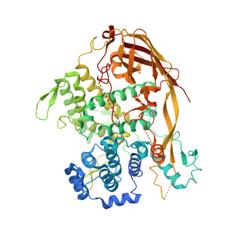
    Plastic waste management is a pressing ecological, social, and economic challenge. The saliva of the lepidopteran Galleria mellonella larvae is capable of oxidizing and depolymerizing polyethylene in hours at room temperature. Here, we analyze by cryo-electron microscopy (cryo-EM) G. mellonella 's saliva directly from the native source. The three-dimensional reconstructions reveal that the buccal secretion is mainly composed of four hexamerins belonging to the hemocyanin/phenoloxidase family, renamed Demetra, Cibeles, Ceres, and a previously unidentified factor termed Cora. Functional assays show that this factor, as its counterparts Demetra and Ceres, is also able to oxidize and degrade polyethylene. The cryo-EM data and the x-ray analysis from purified fractions show that they self-assemble primarily into three macromolecular complexes with striking structural differences that likely modulate their activity. Overall, these results establish the ground to further explore the hexamerins' functionalities, their role in vivo, and their eventual biotechnological application.


  • Organizational Affiliation
    • Department of Structural and Chemical Biology, Centro de Investigaciones Biológicas Margarita Salas, CSIC, 28040 Madrid, Spain.

Macromolecule Content 

  • Total Structure Weight: 502.71 kDa 
  • Atom Count: 33,022 
  • Modeled Residue Count: 3,816 
  • Deposited Residue Count: 4,236 
  • Unique protein chains: 1

Macromolecules

Find similar proteins by:|  3D Structure
Entity ID: 1
MoleculeChains  Sequence LengthOrganismDetailsImage
acidic juvenile hormone-suppressible protein 1
A, B, C, D, E
A, B, C, D, E, F
706Galleria mellonellaMutation(s): 0 

Oligosaccharides

Help  
Entity ID: 2
MoleculeChains Length2D Diagram GlycosylationD Interactions
2-acetamido-2-deoxy-beta-D-glucopyranose-(1-4)-2-acetamido-2-deoxy-beta-D-glucopyranose
G, I, K, M, O
G, I, K, M, O, Q
2N-Glycosylation
Glycosylation Resources
GlyTouCan: G42666HT
GlyCosmos: G42666HT
GlyGen: G42666HT
Entity ID: 3
MoleculeChains Length2D Diagram GlycosylationD Interactions
alpha-D-mannopyranose-(1-2)-alpha-D-mannopyranose-(1-2)-alpha-D-mannopyranose-(1-3)-[alpha-D-mannopyranose-(1-3)-[alpha-D-mannopyranose-(1-6)]alpha-D-mannopyranose-(1-6)]beta-D-mannopyranose-(1-4)-2-acetamido-2-deoxy-beta-D-glucopyranose-(1-4)-2-acetamido-2-deoxy-beta-D-glucopyranose
H, J, L, N, P
H, J, L, N, P, R
9N-Glycosylation
Glycosylation Resources
GlyTouCan: G68668TB
GlyCosmos: G68668TB
GlyGen: G68668TB

Small Molecules

Ligands 1 Unique
IDChains Name / Formula / InChI Key2D Diagram3D Interactions
CU
(Subject of Investigation/LOI)

Query on CU


Download:Ideal Coordinates CCD File
AA [auth E]
BA [auth E]
CA [auth F]
DA [auth F]
S [auth A]
AA [auth E],
BA [auth E],
CA [auth F],
DA [auth F],
S [auth A],
T [auth A],
U [auth B],
V [auth B],
W [auth C],
X [auth C],
Y [auth D],
Z [auth D]
COPPER (II) ION
Cu
JPVYNHNXODAKFH-UHFFFAOYSA-N

Experimental Data & Validation

Experimental Data

  • Method: ELECTRON MICROSCOPY
  • Resolution: 2.85 Å
  • Aggregation State: PARTICLE 
  • Reconstruction Method: SINGLE PARTICLE 
EM Software:
TaskSoftware PackageVersion
RECONSTRUCTIONRELION3.1
MODEL REFINEMENTPHENIX1.19

Structure Validation

View Full Validation Report


Entry History 

& Funding Information

Deposition Data


Funding OrganizationLocationGrant Number
Roechling StiftungGermany--
NATO Science for Peace and Security ProgramBelgiumSPS G5536
Junta de Castilla y Leon, Consejeria de Educacion y Cultura y Fondo Social EuropeoSpainBU263P18
Spanish Ministry of Science, Innovation, and UniversitiesSpainPID2019-111215RB-100
Generalitat de CatalunyaSpain2017 SGR 1192
Spanish Ministry of Science, Innovation, and UniversitiesSpainPID2020-120275GB-I00
Spanish National Research CouncilSpain--

Revision History  (Full details and data files)

  • Version 1.0: 2023-10-04
    Type: Initial release
  • Version 1.1: 2024-11-20
    Changes: Data collection, Structure summary