8C4I | pdb_00008c4i

Ligand-free Crystal Structure of the decameric Sulfofructose Transaldolase BmSF-TAL


Experimental Data Snapshot

  • Method: X-RAY DIFFRACTION
  • Resolution: 3.20 Å
  • R-Value Free: 
    0.268 (Depositor), 0.261 (DCC) 
  • R-Value Work: 
    0.235 (Depositor), 0.234 (DCC) 

Starting Model: experimental
View more details

wwPDB Validation 3D Report Full Report

Validation slider image for 8C4I

This is version 1.3 of the entry. See complete history

Literature

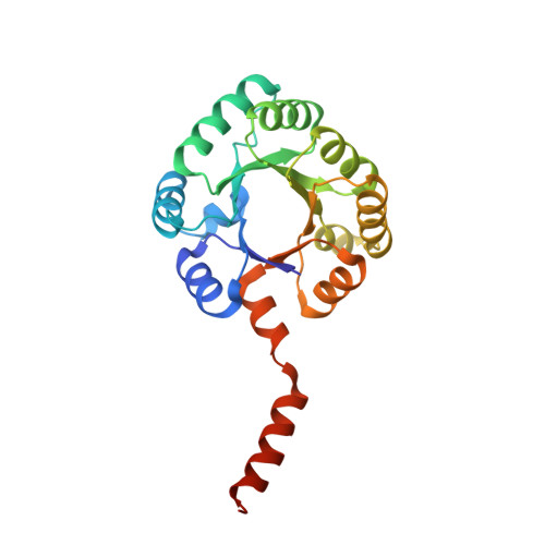
Structure and mechanism of sulfofructose transaldolase, a key enzyme in sulfoquinovose metabolism.

Snow, A.J.D.Sharma, M.Abayakoon, P.Williams, S.J.Blaza, J.N.Davies, G.J.

(2023) Structure 31: 244

  • DOI: https://doi.org/10.1016/j.str.2023.01.010
  • Primary Citation Related Structures: 
    8BC2, 8BC3, 8BC4, 8C4I

  • PubMed Abstract: 

    Sulfoquinovose (SQ) is a key component of plant sulfolipids (sulfoquinovosyl diacylglycerols) and a major environmental reservoir of biological sulfur. Breakdown of SQ is achieved by bacteria through the pathways of sulfoglycolysis. The sulfoglycolytic sulfofructose transaldolase (sulfo-SFT) pathway is used by gut-resident firmicutes and soil saprophytes. After isomerization of SQ to sulfofructose (SF), the namesake enzyme catalyzes the transaldol reaction of SF transferring dihydroxyacetone to 3C/4C acceptors to give sulfolactaldehyde and fructose-6-phosphate or sedoheptulose-7-phosphate. We report the 3D cryo-EM structure of SF transaldolase from Bacillus megaterium in apo and ligand bound forms, revealing a decameric structure formed from two pentameric rings of the protomer. We demonstrate a covalent "Schiff base" intermediate formed by reaction of SF with Lys89 within a conserved Asp-Lys-Glu catalytic triad and defined by an Arg-Trp-Arg sulfonate recognition triad. The structural characterization of the signature enzyme of the sulfo-SFT pathway provides key insights into molecular recognition of the sulfonate group of sulfosugars.


  • Organizational Affiliation
    • Department of Chemistry, York Structural Biology Laboratory, York YO10 5DD, UK.

Macromolecule Content 

  • Total Structure Weight: 253.12 kDa 
  • Atom Count: 16,885 
  • Modeled Residue Count: 2,191 
  • Deposited Residue Count: 2,260 
  • Unique protein chains: 1

Macromolecules

Find similar proteins by:|  3D Structure
Entity ID: 1
MoleculeChains  Sequence LengthOrganismDetailsImage
BmSF-TAL226Priestia aryabhattaiMutation(s): 0 
EC: 2.2.1.14
UniProt
Find proteins for A0A7W3N5X5 (Priestia aryabhattai)
Explore A0A7W3N5X5 
Go to UniProtKB:  A0A7W3N5X5
Entity Groups
Sequence Clusters30% Identity50% Identity70% Identity90% Identity95% Identity100% Identity
UniProt GroupA0A7W3N5X5
Sequence Annotations
Expand
Reference Sequence

Experimental Data & Validation

Experimental Data

  • Method: X-RAY DIFFRACTION
  • Resolution: 3.20 Å
  • R-Value Free:  0.268 (Depositor), 0.261 (DCC) 
  • R-Value Work:  0.235 (Depositor), 0.234 (DCC) 
Space Group: P 1 21 1
Unit Cell:
Length ( Å )Angle ( ˚ )
a = 73.891α = 90
b = 120.839β = 93.401
c = 152.643γ = 90
Software Package:
Software NamePurpose
REFMACrefinement
REFMACrefinement
Aimlessdata scaling
MOLREPphasing

Structure Validation

View Full Validation Report


Entry History 

& Funding Information

Deposition Data


Funding OrganizationLocationGrant Number
Leverhulme TrustUnited KingdomRPG-2017-190

Revision History  (Full details and data files)

  • Version 1.0: 2023-02-22
    Type: Initial release
  • Version 1.1: 2023-03-08
    Changes: Database references
  • Version 1.2: 2023-03-15
    Changes: Database references
  • Version 1.3: 2023-11-15
    Changes: Data collection, Source and taxonomy