8C3H

Cereblon isoform 4 from Magnetospirillum gryphiswaldense in complex a long aspartimide degron peptide


Experimental Data Snapshot

  • Method: X-RAY DIFFRACTION
  • Resolution: 1.71 Å
  • R-Value Free: 0.210 
  • R-Value Work: 0.179 

wwPDB Validation   3D Report Full Report


This is version 2.0 of the entry. See complete history


Literature

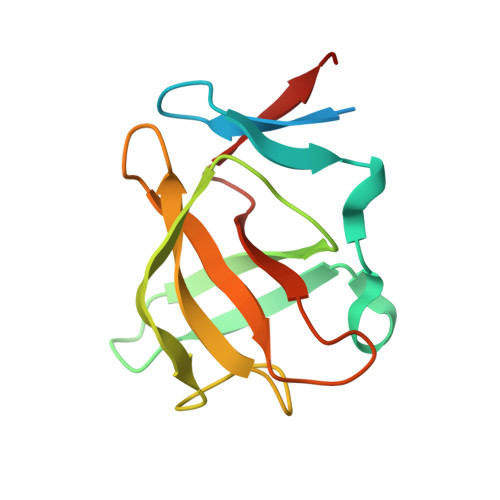
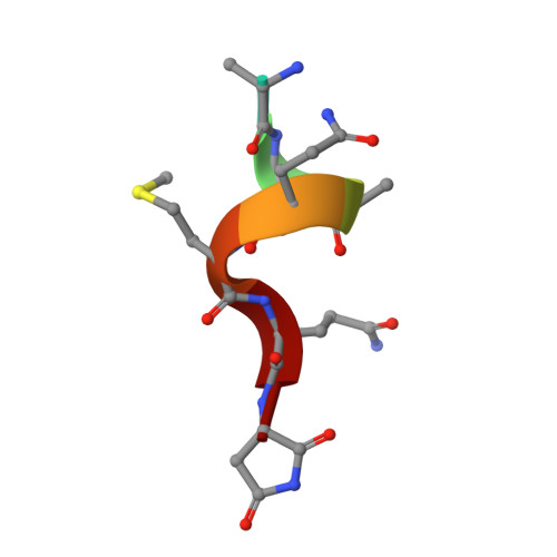
Cereblon neo-substrate binding mimics the recognition of the cyclic imide degron.

Heim, C.Spring, A.K.Kirchgassner, S.Schwarzer, D.Hartmann, M.D.

(2023) Biochem Biophys Res Commun 646: 30-35

  • DOI: https://doi.org/10.1016/j.bbrc.2023.01.051
  • Primary Citation of Related Structures:  
    8C3H

  • PubMed Abstract: 

    In targeted protein degradation, immunomodulatory drugs (IMiDs) or cereblon (CRBN) E3 ligase modulatory drugs (CELMoDs) recruit neo-substrate proteins to the E3 ubiquitin ligase receptor CRBN for ubiquitination and subsequent proteasomal degradation. While the structural basis of this mechanism is generally understood, we have only recently described the recognition mode of the natural CRBN degron. In this communication, we reveal that the IMiD- or CELMoD-mediated binding of neo-substrates closely mimics the recognition of natural degrons. In crystal structures, we identify a conserved binding mode for natural degron peptides with an elaborate hydrogen bonding network involving the backbone of each of the six C-terminal degron residues, without the involvement of side chains. In a structural comparison, we show that neo-substrates recruited by IMiDs or CELMoDs emulate every single hydrogen bond of this network and thereby explain the origins of the largely sequence-independent recognition of neo-substrates. Our results imply that the V388I substitution in CRBN does not impair natural degron recognition and complete the structural basis for the rational design of CRBN effectors.


  • Organizational Affiliation

    Max Planck Institute for Biology, Tübingen, Germany; Interfaculty Institute of Biochemistry, University of Tübingen, Tübingen, Germany; NanoTemper Technologies GmbH, Munich, Germany.


Macromolecules
Find similar proteins by:  (by identity cutoff)  |  3D Structure
Entity ID: 1
MoleculeChains Sequence LengthOrganismDetailsImage
Cereblon isoform 4
A, B, C
124Magnetospirillum gryphiswaldenseMutation(s): 0 
Gene Names: MGR_0879
UniProt
Find proteins for A4TVL0 (Magnetospirillum gryphiswaldense)
Explore A4TVL0 
Go to UniProtKB:  A4TVL0
Entity Groups  
Sequence Clusters30% Identity50% Identity70% Identity90% Identity95% Identity100% Identity
UniProt GroupA4TVL0
Sequence Annotations
Expand
  • Reference Sequence

Find similar proteins by:  Sequence   |   3D Structure  

Entity ID: 2
MoleculeChains Sequence LengthOrganismDetailsImage
P3(40)
D, E
8Homo sapiensMutation(s): 0 
UniProt & NIH Common Fund Data Resources
Find proteins for P05067 (Homo sapiens)
Explore P05067 
Go to UniProtKB:  P05067
PHAROS:  P05067
GTEx:  ENSG00000142192 
Entity Groups  
UniProt GroupP05067
Sequence Annotations
Expand
  • Reference Sequence
Small Molecules
Experimental Data & Validation

Experimental Data

  • Method: X-RAY DIFFRACTION
  • Resolution: 1.71 Å
  • R-Value Free: 0.210 
  • R-Value Work: 0.179 
  • Space Group: P 21 21 21
Unit Cell:
Length ( Å )Angle ( ˚ )
a = 56.256α = 90
b = 58.821β = 90
c = 88.858γ = 90
Software Package:
Software NamePurpose
REFMACrefinement
XDSdata reduction
XDSdata scaling

Structure Validation

View Full Validation Report


Entry History & Funding Information

Deposition Data


Funding OrganizationLocationGrant Number
Not funded--

Revision History  (Full details and data files)

  • Version 1.0: 2023-02-08
    Type: Initial release
  • Version 2.0: 2023-11-15
    Changes: Advisory, Atomic model, Data collection, Derived calculations, Refinement description