Fast viral dynamics revealed by microsecond time-resolved cryo-EM.
Harder, O.F., Barrass, S.V., Drabbels, M., Lorenz, U.J.(2023) Nat Commun 14: 5649-5649
- PubMed: 37704664 Search on PubMedSearch on PubMed Central
- DOI: https://doi.org/10.1038/s41467-023-41444-x
- Primary Citation Related Structures:
8C38, 8CPY - PubMed Abstract:
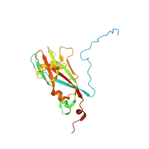
Observing proteins as they perform their tasks has largely remained elusive, which has left our understanding of protein function fundamentally incomplete. To enable such observations, we have recently proposed a technique that improves the time resolution of cryo-electron microscopy (cryo-EM) to microseconds. Here, we demonstrate that microsecond time-resolved cryo-EM enables observations of fast protein dynamics. We use our approach to elucidate the mechanics of the capsid of cowpea chlorotic mottle virus (CCMV), whose large-amplitude motions play a crucial role in the viral life cycle. We observe that a pH jump causes the extended configuration of the capsid to contract on the microsecond timescale. While this is a concerted process, the motions of the capsid proteins involve different timescales, leading to a curved reaction path. It is difficult to conceive how such a detailed picture of the dynamics could have been obtained with any other method, which highlights the potential of our technique. Crucially, our experiments pave the way for microsecond time-resolved cryo-EM to be applied to a broad range of protein dynamics that previously could not have been observed. This promises to fundamentally advance our understanding of protein function.
- Ecole Polytechnique Fédérale de Lausanne (EPFL), Laboratory of Molecular Nanodynamics, CH-1015, Lausanne, Switzerland.
Organizational Affiliation: