8C07 | pdb_00008c07

Structure of HECT E3 UBR5 forming K48 linked Ubiquitin chains


Experimental Data Snapshot

  • Method: ELECTRON MICROSCOPY
  • Resolution: 3.30 Å
  • Aggregation State: PARTICLE 
  • Reconstruction Method: SINGLE PARTICLE 

wwPDB Validation 3D Report Full Report

Validation slider image for 8C07

This is version 1.4 of the entry. See complete history

Literature

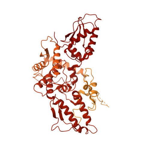
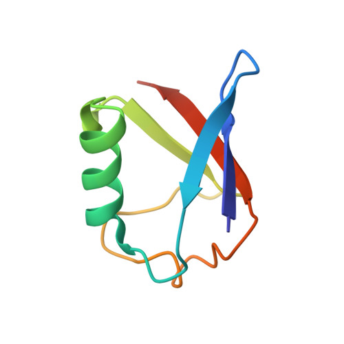
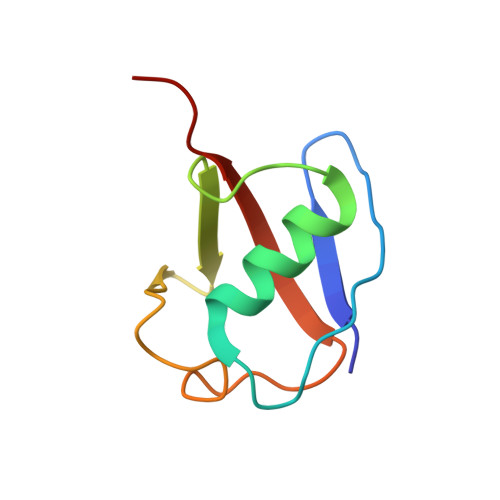
Structural snapshots along K48-linked ubiquitin chain formation by the HECT E3 UBR5.

Hehl, L.A.Horn-Ghetko, D.Prabu, J.R.Vollrath, R.Vu, D.T.Perez Berrocal, D.A.Mulder, M.P.C.van der Heden van Noort, G.J.Schulman, B.A.

(2024) Nat Chem Biol 20: 190-200

  • DOI: https://doi.org/10.1038/s41589-023-01414-2
  • Primary Citation Related Structures: 
    8C06, 8C07

  • PubMed Abstract: 

    Ubiquitin (Ub) chain formation by homologous to E6AP C-terminus (HECT)-family E3 ligases regulates vast biology, yet the structural mechanisms remain unknown. We used chemistry and cryo-electron microscopy (cryo-EM) to visualize stable mimics of the intermediates along K48-linked Ub chain formation by the human E3, UBR5. The structural data reveal a ≈ 620 kDa UBR5 dimer as the functional unit, comprising a scaffold with flexibly tethered Ub-associated (UBA) domains, and elaborately arranged HECT domains. Chains are forged by a UBA domain capturing an acceptor Ub, with its K48 lured into the active site by numerous interactions between the acceptor Ub, manifold UBR5 elements and the donor Ub. The cryo-EM reconstructions allow defining conserved HECT domain conformations catalyzing Ub transfer from E2 to E3 and from E3. Our data show how a full-length E3, ubiquitins to be adjoined, E2 and intermediary products guide a feed-forward HECT domain conformational cycle establishing a highly efficient, broadly targeting, K48-linked Ub chain forging machine.


  • Organizational Affiliation
    • Department of Chemistry, School of Natural Sciences, Technical University of Munich, Garching, Germany.

Macromolecule Content 

  • Total Structure Weight: 637.76 kDa 
  • Atom Count: 4,974 
  • Modeled Residue Count: 644 
  • Deposited Residue Count: 5,764 
  • Unique protein chains: 3

Macromolecules

Find similar proteins by:|  3D Structure
Entity ID: 1
MoleculeChains  Sequence LengthOrganismDetailsImage
E3 ubiquitin-protein ligase UBR5A [auth B],
D [auth J]
2,806Homo sapiensMutation(s): 2 
Gene Names: UBR5EDDEDD1HYDKIAA0896
EC: 2.3.2.26
UniProt & NIH Common Fund Data Resources
Find proteins for O95071 (Homo sapiens)
Explore O95071 
Go to UniProtKB:  O95071
PHAROS:  O95071
GTEx:  ENSG00000104517 
Entity Groups
Sequence Clusters30% Identity50% Identity70% Identity90% Identity95% Identity100% Identity
UniProt GroupO95071
Sequence Annotations
Expand
Reference Sequence
Find similar proteins by:|  3D Structure
Entity ID: 2
MoleculeChains  Sequence LengthOrganismDetailsImage
Polyubiquitin-BB [auth I]76Homo sapiensMutation(s): 1 
Gene Names: UBB
UniProt & NIH Common Fund Data Resources
Find proteins for P0CG47 (Homo sapiens)
Explore P0CG47 
Go to UniProtKB:  P0CG47
PHAROS:  P0CG47
GTEx:  ENSG00000170315 
Entity Groups
Sequence Clusters30% Identity50% Identity70% Identity90% Identity95% Identity100% Identity
UniProt GroupP0CG47
Sequence Annotations
Expand
Reference Sequence
Find similar proteins by:|  3D Structure
Entity ID: 3
MoleculeChains  Sequence LengthOrganismDetailsImage
Polyubiquitin-BC [auth K]76Homo sapiensMutation(s): 0 
Gene Names: UBB
UniProt & NIH Common Fund Data Resources
Find proteins for P0CG47 (Homo sapiens)
Explore P0CG47 
Go to UniProtKB:  P0CG47
PHAROS:  P0CG47
GTEx:  ENSG00000170315 
Entity Groups
Sequence Clusters30% Identity50% Identity70% Identity90% Identity95% Identity100% Identity
UniProt GroupP0CG47
Sequence Annotations
Expand
Reference Sequence

Small Molecules

Ligands 1 Unique
IDChains Name / Formula / InChI Key2D Diagram3D Interactions
SY8

Query on SY8


Download:Ideal Coordinates CCD File
E [auth K]5-azanylpentan-2-one
C5 H11 N O
VTHKLCWOLNQWHB-UHFFFAOYSA-N

Experimental Data & Validation

Experimental Data

  • Method: ELECTRON MICROSCOPY
  • Resolution: 3.30 Å
  • Aggregation State: PARTICLE 
  • Reconstruction Method: SINGLE PARTICLE 
EM Software:
TaskSoftware PackageVersion
MODEL REFINEMENTPHENIX

Structure Validation

View Full Validation Report


Entry History 

& Funding Information

Deposition Data


Funding OrganizationLocationGrant Number
Max Planck SocietyGermany--

Revision History  (Full details and data files)

  • Version 1.0: 2023-08-23
    Type: Initial release
  • Version 1.1: 2023-09-06
    Changes: Database references
  • Version 1.2: 2024-02-14
    Changes: Database references
  • Version 1.3: 2024-10-09
    Changes: Data collection, Structure summary
  • Version 1.4: 2025-07-09
    Changes: Data collection