8BWF

PTBP1 RRM1 bound to an allosteric inhibitor


Experimental Data Snapshot

  • Method: X-RAY DIFFRACTION
  • Resolution: 2.90 Å
  • R-Value Free: 0.340 
  • R-Value Work: 0.243 
  • R-Value Observed: 0.248 

wwPDB Validation   3D Report Full Report


This is version 2.0 of the entry. See complete history


Literature

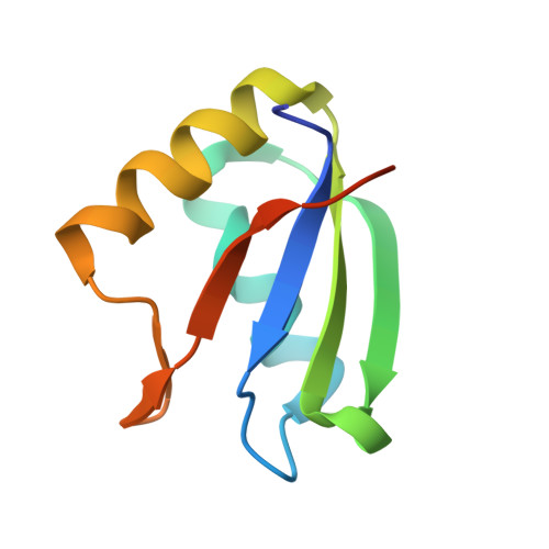
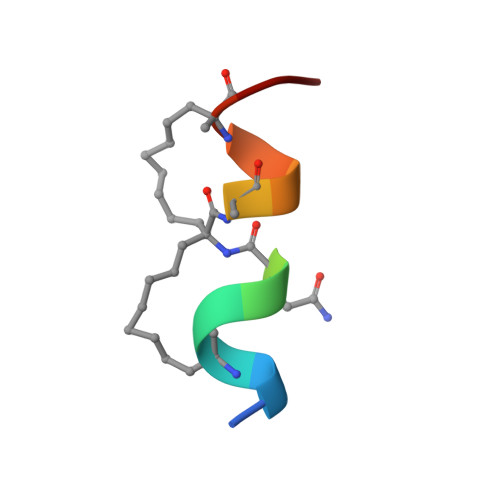
Rationally designed stapled peptides allosterically inhibit PTBP1-RNA-binding.

Schmeing, S.Amrahova, G.Bigler, K.Chang, J.Y.Openy, J.Pal, S.Posada, L.Gasper, R.'t Hart, P.

(2023) Chem Sci 14: 8269-8278

  • DOI: https://doi.org/10.1039/d3sc00985h
  • Primary Citation of Related Structures:  
    8BWF

  • PubMed Abstract: 

    The diverse role of the splicing factor PTBP1 in human cells has been widely studied and was found to be a driver for several diseases. PTBP1 binds RNA through its RNA-recognition motifs which lack obvious pockets for inhibition. A unique transient helix has been described to be part of its first RNA-recognition motif and to be important for RNA binding. In this study, we further confirmed the role of this helix and envisioned its dynamic nature as a unique opportunity to develop stapled peptide inhibitors of PTBP1. The peptides were found to be able to inhibit RNA binding via fluorescence polarization assays and directly occupy the helix binding site as observed by protein crystallography. These cell-permeable inhibitors were validated in cellulo to alter the regulation of alternative splicing events regulated by PTBP1. Our study demonstrates transient secondary structures of a protein can be mimicked by stapled peptides to inhibit allosteric mechanisms.


  • Organizational Affiliation

    Chemical Genomics Centre of the Max Planck Society, Max Planck Institute of Molecular Physiology Otto-Hahn-Strasse 11 44227 Dortmund Germany peter.t-hart@mpi-dortmund.mpg.de.


Macromolecules
Find similar proteins by:  (by identity cutoff)  |  3D Structure
Entity ID: 1
MoleculeChains Sequence LengthOrganismDetailsImage
Polypyrimidine tract-binding protein 186Homo sapiensMutation(s): 0 
Gene Names: PTBP1PTB
UniProt & NIH Common Fund Data Resources
Find proteins for P26599 (Homo sapiens)
Explore P26599 
Go to UniProtKB:  P26599
PHAROS:  P26599
GTEx:  ENSG00000011304 
Entity Groups  
Sequence Clusters30% Identity50% Identity70% Identity90% Identity95% Identity100% Identity
UniProt GroupP26599
Sequence Annotations
Expand
  • Reference Sequence

Find similar proteins by:  Sequence   |   3D Structure  

Entity ID: 2
MoleculeChains Sequence LengthOrganismDetailsImage
Ligand13Homo sapiensMutation(s): 0 
Entity Groups  
Sequence Clusters30% Identity50% Identity70% Identity90% Identity95% Identity100% Identity
Sequence Annotations
Expand
  • Reference Sequence
Small Molecules
Ligands 3 Unique
IDChains Name / Formula / InChI Key2D Diagram3D Interactions
SO4
Query on SO4

Download Ideal Coordinates CCD File 
BB [auth g]
CB [auth H]
GA [auth A]
HA [auth A]
IA [auth A]
BB [auth g],
CB [auth H],
GA [auth A],
HA [auth A],
IA [auth A],
IB [auth K],
JA [auth A],
JB [auth K],
KA [auth A],
LB [auth L],
NA [auth C],
PB [auth O],
QA [auth D],
RA [auth D],
UA [auth F],
VA [auth F],
YA [auth G],
ZA [auth G]
SULFATE ION
O4 S
QAOWNCQODCNURD-UHFFFAOYSA-L
GOL
Query on GOL

Download Ideal Coordinates CCD File 
EB [auth I],
GB [auth i],
OA [auth C],
WA [auth F]
GLYCEROL
C3 H8 O3
PEDCQBHIVMGVHV-UHFFFAOYSA-N
NH2
Query on NH2

Download Ideal Coordinates CCD File 
AB [auth g]
DB [auth h]
FB [auth i]
HB [auth j]
KB [auth k]
AB [auth g],
DB [auth h],
FB [auth i],
HB [auth j],
KB [auth k],
LA [auth a],
MA [auth b],
MB [auth l],
NB [auth m],
OB [auth n],
PA [auth c],
QB [auth o],
RB [auth p],
SA [auth d],
TA [auth e],
XA [auth f]
AMINO GROUP
H2 N
QGZKDVFQNNGYKY-UHFFFAOYSA-N
Modified Residues  1 Unique
IDChains TypeFormula2D DiagramParent
MK8
Query on MK8
B [auth a]
BA [auth n]
D [auth b]
DA [auth o]
F [auth c]
B [auth a],
BA [auth n],
D [auth b],
DA [auth o],
F [auth c],
FA [auth p],
H [auth d],
J [auth e],
L [auth f],
N [auth g],
P [auth h],
R [auth i],
T [auth j],
V [auth k],
X [auth l],
Z [auth m]
L-PEPTIDE LINKINGC7 H15 N O2LEU
Experimental Data & Validation

Experimental Data

  • Method: X-RAY DIFFRACTION
  • Resolution: 2.90 Å
  • R-Value Free: 0.340 
  • R-Value Work: 0.243 
  • R-Value Observed: 0.248 
  • Space Group: P 21 21 2
Unit Cell:
Length ( Å )Angle ( ˚ )
a = 244.2α = 90
b = 76.63β = 90
c = 94.13γ = 90
Software Package:
Software NamePurpose
PHENIXrefinement
XSCALEdata scaling
XDSdata reduction
PHASERphasing

Structure Validation

View Full Validation Report


Entry History & Funding Information

Deposition Data


Funding OrganizationLocationGrant Number
Other private--

Revision History  (Full details and data files)

  • Version 1.0: 2023-10-11
    Type: Initial release
  • Version 2.0: 2023-11-15
    Changes: Atomic model, Data collection, Derived calculations