8BV9 | pdb_00008bv9

Acylphosphatase from E. coli


Experimental Data Snapshot

  • Method: X-RAY DIFFRACTION
  • Resolution: 2.55 Å
  • R-Value Free: 
    0.278 (Depositor), 0.291 (DCC) 
  • R-Value Work: 
    0.205 (Depositor), 0.214 (DCC) 

Starting Model: experimental
View more details

wwPDB Validation   3D Report Full Report


This is version 2.0 of the entry. See complete history


Literature

First 3-D structural evidence of a native-like intertwined dimer in the acylphosphatase family.

Martinez-Rodriguez, S.Camara-Artigas, A.Gavira, J.A.

(2023) Biochem Biophys Res Commun 682: 85-90

  • DOI: https://doi.org/10.1016/j.bbrc.2023.09.053
  • Primary Citation of Related Structures:  
    8BV9

  • PubMed Abstract: 

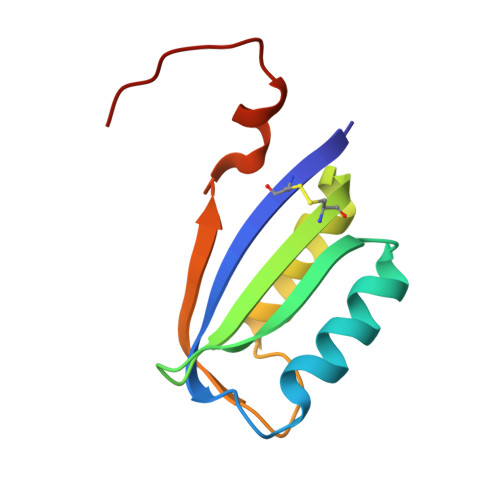
    Acylphosphatase (AcP, EC 3.6.1.7) is a small model protein conformed by a ferredoxin-like fold, profoundly studied to get insights into protein folding and aggregation processes. Numerous studies focused on the aggregation and/or amyloidogenic properties of AcPs suggest the importance of edge-β-strands in the process. In this work, we present the first crystallographic structure of Escherichia coli AcP (EcoAcP), showing notable differences with the only available NMR structure for this enzyme. EcoAcP is crystalised as an intertwined dimer formed by replacing a single C-terminal β-strand between two protomers, suggesting a flexible character of the C-terminal edge of EcoAcP. Despite numerous works where AcP from different sources have been used as a model system for protein aggregation, our domain-swapped EcoAcP structure is the first 3-D structural evidence of native-like aggregated species for any AcP reported to date, providing clues on molecular determinants unleashing aggregation.


  • Organizational Affiliation
    • Department of Biochemistry and Molecular Biology III and Immunology, University of Granada, Avenida de La Investigación 11, Granada, 18071, Spain; Laboratorio de Estudios Cristalográficos, CSIC-UGR, Avda. de Las Palmeras 4, Armilla, Granada, 18100, Spain. Electronic address: sergio@ugr.es.

Macromolecules
Find similar proteins by:  (by identity cutoff)  |  3D Structure
Entity ID: 1
MoleculeChains Sequence LengthOrganismDetailsImage
Acylphosphatase93Escherichia coliMutation(s): 0 
Gene Names: yccXZ1320ECs1052
EC: 3.6.1.7
UniProt
Find proteins for P0AB65 (Escherichia coli (strain K12))
Explore P0AB65 
Go to UniProtKB:  P0AB65
Entity Groups  
Sequence Clusters30% Identity50% Identity70% Identity90% Identity95% Identity100% Identity
UniProt GroupP0AB65
Sequence Annotations
Expand
  • Reference Sequence
Experimental Data & Validation

Experimental Data

  • Method: X-RAY DIFFRACTION
  • Resolution: 2.55 Å
  • R-Value Free:  0.278 (Depositor), 0.291 (DCC) 
  • R-Value Work:  0.205 (Depositor), 0.214 (DCC) 
Space Group: C 2 2 2
Unit Cell:
Length ( Å )Angle ( ˚ )
a = 40.415α = 90
b = 81.944β = 90
c = 63.994γ = 90
Software Package:
Software NamePurpose
REFMACrefinement
autoPROCdata reduction
Aimlessdata scaling
PHENIXphasing

Structure Validation

View Full Validation Report


Entry History & Funding Information

Deposition Data


Funding OrganizationLocationGrant Number
Ministerio de Ciencia e Innovacion (MCIN)SpainPID2020-116261GB-I00

Revision History  (Full details and data files)

  • Version 1.0: 2023-10-18
    Type: Initial release
  • Version 1.1: 2023-10-25
    Changes: Database references
  • Version 1.2: 2024-10-23
    Changes: Structure summary
  • Version 2.0: 2025-11-05
    Type: Coordinate replacement
    Reason: Chirality error
    Changes: Advisory, Atomic model, Data collection, Database references, Derived calculations, Non-polymer description, Other, Polymer sequence, Refinement description, Source and taxonomy, Structure summary