8BSZ | pdb_00008bsz

Notum Inhibitor ARUK3005522


Experimental Data Snapshot

  • Method: X-RAY DIFFRACTION
  • Resolution: 1.70 Å
  • R-Value Free: 
    0.220 (Depositor), 0.221 (DCC) 
  • R-Value Work: 
    0.196 (Depositor), 0.196 (DCC) 
  • R-Value Observed: 
    0.197 (Depositor) 

Starting Model: experimental
View more details

wwPDB Validation 3D Report Full Report

Validation slider image for 8BSZ

Ligand Structure Quality Assessment 


This is version 1.3 of the entry. See complete history

Literature

Designed switch from covalent to non-covalent inhibitors of carboxylesterase Notum activity.

Atkinson, B.N.Willis, N.J.Zhao, Y.Patel, C.Frew, S.Costelloe, K.Magno, L.Svensson, F.Jones, E.Y.Fish, P.V.

(2023) Eur J Med Chem 251: 115132-115132

  • DOI: https://doi.org/10.1016/j.ejmech.2023.115132
  • Primary Citation Related Structures: 
    8BSP, 8BSQ, 8BSR, 8BSZ, 8BT0, 8BT2, 8BT5, 8BT7, 8BT8, 8BTA, 8BTC, 8BTE, 8BTH, 8BTI

  • PubMed Abstract: 

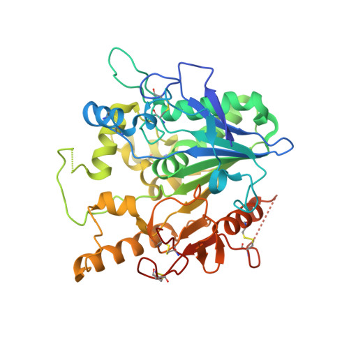
    N-Acyl indolines 4 are potent, non-covalent Notum inhibitors developed from a covalent virtual screening hit 2a. The lead compounds were simple to synthesise, achieved excellent potency in a biochemical Notum-OPTS assay and restored Wnt signalling in a cell-based TCF/LEF reporter assay. Multiple high resolution X-ray structures established a common binding mode of these inhibitors with the indoline bound centred in the palmiteolate pocket with key interactions being aromatic stacking and a water mediated hydrogen bond to the oxyanion hole. These N-acyl indolines 4 will be useful tools for use in vitro studies to investigate the role of Notum in disease models, especially when paired with a structurally related covalent inhibitor (e.g. 4w and 2a). Overall, this study highlights the designed switch from covalent to non-covalent Notum inhibitors and so illustrates a complementary approach for hit generation and target inhibition.


  • Organizational Affiliation
    • Alzheimer's Research UK UCL Drug Discovery Institute, University College London, The Cruciform Building, Gower Street, London, WC1E 6BT, UK.

Macromolecule Content 

  • Total Structure Weight: 44.8 kDa 
  • Atom Count: 3,066 
  • Modeled Residue Count: 358 
  • Deposited Residue Count: 383 
  • Unique protein chains: 1

Macromolecules

Find similar proteins by:|  3D Structure
Entity ID: 1
MoleculeChains  Sequence LengthOrganismDetailsImage
Palmitoleoyl-protein carboxylesterase NOTUM383Homo sapiensMutation(s): 1 
Gene Names: NOTUMOK/SW-CL.30
EC: 3.1.1.98
UniProt & NIH Common Fund Data Resources
Find proteins for Q6P988 (Homo sapiens)
Explore Q6P988 
Go to UniProtKB:  Q6P988
PHAROS:  Q6P988
GTEx:  ENSG00000185269 
Entity Groups
Sequence Clusters30% Identity50% Identity70% Identity90% Identity95% Identity100% Identity
UniProt GroupQ6P988
Glycosylation
Glycosylation Sites: 1Go to GlyGen: Q6P988-1
Sequence Annotations
Expand
Reference Sequence

Small Molecules

Ligands 5 Unique
IDChains Name / Formula / InChI Key2D Diagram3D Interactions
NAG

Query on NAG


Download:Ideal Coordinates CCD File
B [auth A]2-acetamido-2-deoxy-beta-D-glucopyranose
C8 H15 N O6
OVRNDRQMDRJTHS-FMDGEEDCSA-N
RGU
(Subject of Investigation/LOI)

Query on RGU


Download:Ideal Coordinates CCD File
M [auth A](3~{a}~{R})-2,3,3~{a},4-tetrahydropyrrolo[1,2-a]indol-1-one
C11 H11 N O
QPPMCKRHXDXQPA-SECBINFHSA-N
SO4

Query on SO4


Download:Ideal Coordinates CCD File
C [auth A],
D [auth A],
E [auth A],
N [auth A]
SULFATE ION
O4 S
QAOWNCQODCNURD-UHFFFAOYSA-L
DMS

Query on DMS


Download:Ideal Coordinates CCD File
F [auth A]DIMETHYL SULFOXIDE
C2 H6 O S
IAZDPXIOMUYVGZ-UHFFFAOYSA-N
EDO

Query on EDO


Download:Ideal Coordinates CCD File
G [auth A]
H [auth A]
I [auth A]
J [auth A]
K [auth A]
G [auth A],
H [auth A],
I [auth A],
J [auth A],
K [auth A],
L [auth A]
1,2-ETHANEDIOL
C2 H6 O2
LYCAIKOWRPUZTN-UHFFFAOYSA-N

Experimental Data & Validation

Experimental Data

  • Method: X-RAY DIFFRACTION
  • Resolution: 1.70 Å
  • R-Value Free:  0.220 (Depositor), 0.221 (DCC) 
  • R-Value Work:  0.196 (Depositor), 0.196 (DCC) 
  • R-Value Observed: 0.197 (Depositor) 
Space Group: P 21 21 21
Unit Cell:
Length ( Å )Angle ( ˚ )
a = 59.672α = 90
b = 71.414β = 90
c = 78.078γ = 90
Software Package:
Software NamePurpose
PHENIXrefinement
xia2data reduction
xia2data scaling
MOLREPphasing

Structure Validation

View Full Validation Report


Ligand Structure Quality Assessment 


Entry History 

& Funding Information

Deposition Data


Funding OrganizationLocationGrant Number
Cancer Research UKUnited KingdomC375/A17721

Revision History  (Full details and data files)

  • Version 1.0: 2022-12-14
    Type: Initial release
  • Version 1.1: 2023-03-22
    Changes: Refinement description
  • Version 1.2: 2023-06-28
    Changes: Database references
  • Version 1.3: 2024-11-13
    Changes: Data collection, Structure summary