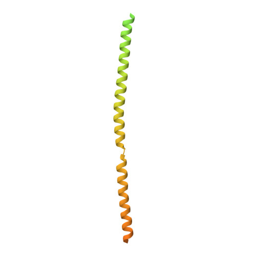
8BS7 | pdb_00008bs7

Multimerisation domain of Borna disease virus 1


Experimental Data Snapshot

  • Method: X-RAY DIFFRACTION
  • Resolution: 3.20 Å
  • R-Value Free: 
    0.387 (Depositor), 0.379 (DCC) 
  • R-Value Work: 
    0.335 (Depositor), 0.337 (DCC) 
  • R-Value Observed: 
    0.338 (Depositor) 

Starting Model: in silico
View more details

wwPDB Validation 3D Report Full Report

Validation slider image for 8BS7

This is version 1.1 of the entry. See complete history

Literature

Structural and biophysical characterization of the Borna disease virus 1 phosphoprotein.

Whitehead, J.D.Grimes, J.M.Keown, J.R.

(2023) Acta Crystallogr F Struct Biol Commun 79: 51-60

  • DOI: https://doi.org/10.1107/S2053230X23000717
  • Primary Citation Related Structures: 
    8BS7

  • PubMed Abstract: 

    Bornaviruses are RNA viruses with a mammalian, reptilian, and avian host range. The viruses infect neuronal cells and in rare cases cause a lethal encephalitis. The family Bornaviridae are part of the Mononegavirales order of viruses, which contain a nonsegmented viral genome. Mononegavirales encode a viral phosphoprotein (P) that binds both the viral polymerase (L) and the viral nucleoprotein (N). The P protein acts as a molecular chaperone and is required for the formation of a functional replication/transcription complex. In this study, the structure of the oligomerization domain of the phosphoprotein determined by X-ray crystallography is reported. The structural results are complemented with biophysical characterization using circular dichroism, differential scanning calorimetry and small-angle X-ray scattering. The data reveal the phosphoprotein to assemble into a stable tetramer, with the regions outside the oligomerization domain remaining highly flexible. A helix-breaking motif is observed between the α-helices at the midpoint of the oligomerization domain that appears to be conserved across the Bornaviridae. These data provide information on an important component of the bornavirus replication complex.


  • Organizational Affiliation
    • Division of Structural Biology, Wellcome Trust Centre for Human Genetics, University of Oxford, Oxford, United Kingdom.

Macromolecule Content 

  • Total Structure Weight: 196.83 kDa 
  • Atom Count: 4,525 
  • Modeled Residue Count: 589 
  • Deposited Residue Count: 1,736 
  • Unique protein chains: 1

Macromolecules

Find similar proteins by:|  3D Structure
Entity ID: 1
MoleculeChains  Sequence LengthOrganismDetailsImage
Phosphoprotein
A, B, C, D, E
A, B, C, D, E, F, G, H
217Borna disease virus 1Mutation(s): 0 
Gene Names: P/X
UniProt
Find proteins for P0C799 (Borna disease virus (strain V))
Explore P0C799 
Go to UniProtKB:  P0C799
Entity Groups
Sequence Clusters30% Identity50% Identity70% Identity90% Identity95% Identity100% Identity
UniProt GroupP0C799
Sequence Annotations
Expand
Reference Sequence

Experimental Data & Validation

Experimental Data

  • Method: X-RAY DIFFRACTION
  • Resolution: 3.20 Å
  • R-Value Free:  0.387 (Depositor), 0.379 (DCC) 
  • R-Value Work:  0.335 (Depositor), 0.337 (DCC) 
  • R-Value Observed: 0.338 (Depositor) 
Space Group: P 1
Unit Cell:
Length ( Å )Angle ( ˚ )
a = 34.873α = 89.97
b = 35.154β = 90.01
c = 153.125γ = 90.54
Software Package:
Software NamePurpose
PHENIXrefinement
autoPROCdata scaling
PHASERphasing
autoPROCdata reduction

Structure Validation

View Full Validation Report


Entry History 

& Funding Information

Deposition Data


Funding OrganizationLocationGrant Number
Wellcome TrustUnited Kingdom200835/Z/16/Z

Revision History  (Full details and data files)

  • Version 1.0: 2023-04-05
    Type: Initial release
  • Version 1.1: 2024-06-19
    Changes: Data collection