8BP1 | pdb_00008bp1

Crystal structure of BHMeHis1.0, an engineered enzyme for the Morita-Baylis-Hillman reaction

  • Classification: BIOSYNTHETIC PROTEIN
  • Organism(s): synthetic construct
  • Expression System: Escherichia coli
  • Mutation(s): No 

  • Deposited: 2022-11-15 Released: 2024-02-28 
  • Deposition Author(s): Hardy, F.J.
  • Funding Organization(s): Biotechnology and Biological Sciences Research Council (BBSRC), European Research Council (ERC), Human Frontier Science Program (HFSP)

Experimental Data Snapshot

  • Method: X-RAY DIFFRACTION
  • Resolution: 1.72 Å
  • R-Value Free: 
    0.202 (Depositor), 0.175 (DCC) 
  • R-Value Work: 
    0.181 (Depositor) 
  • R-Value Observed: 
    0.182 (Depositor) 

Starting Model: experimental
View more details

wwPDB Validation 3D Report Full Report

Validation slider image for 8BP1

This is version 1.3 of the entry. See complete history

Literature

A non-canonical nucleophile unlocks a new mechanistic pathway in a designed enzyme.

Hutton, A.E.Foster, J.Crawshaw, R.Hardy, F.J.Johannissen, L.O.Lister, T.M.Gerard, E.F.Birch-Price, Z.Obexer, R.Hay, S.Green, A.P.

(2024) Nat Commun 15: 1956-1956

  • DOI: https://doi.org/10.1038/s41467-024-46123-z
  • Primary Citation Related Structures: 
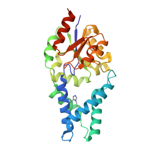
    8BP0, 8BP1

  • PubMed Abstract: 

    Directed evolution of computationally designed enzymes has provided new insights into the emergence of sophisticated catalytic sites in proteins. In this regard, we have recently shown that a histidine nucleophile and a flexible arginine can work in synergy to accelerate the Morita-Baylis-Hillman (MBH) reaction with unrivalled efficiency. Here, we show that replacing the catalytic histidine with a non-canonical N δ -methylhistidine (MeHis23) nucleophile leads to a substantially altered evolutionary outcome in which the catalytic Arg124 has been abandoned. Instead, Glu26 has emerged, which mediates a rate-limiting proton transfer step to deliver an enzyme (BH MeHis 1.8) that is more than an order of magnitude more active than our earlier MBHase. Interestingly, although MeHis23 to His substitution in BH MeHis 1.8 reduces activity by 4-fold, the resulting His containing variant is still a potent MBH biocatalyst. However, analysis of the BH MeHis 1.8 evolutionary trajectory reveals that the MeHis nucleophile was crucial in the early stages of engineering to unlock the new mechanistic pathway. This study demonstrates how even subtle perturbations to key catalytic elements of designed enzymes can lead to vastly different evolutionary outcomes, resulting in new mechanistic solutions to complex chemical transformations.


  • Organizational Affiliation
    • Manchester Institute of Biotechnology, School of Chemistry, The University of Manchester, Manchester, UK.

Macromolecule Content 

  • Total Structure Weight: 28.01 kDa 
  • Atom Count: 2,170 
  • Modeled Residue Count: 231 
  • Deposited Residue Count: 242 
  • Unique protein chains: 1

Macromolecules

Find similar proteins by:|  3D Structure
Entity ID: 1
MoleculeChains  Sequence LengthOrganismDetailsImage
BHMeHis1.0242synthetic constructMutation(s): 0 

Small Molecules

Ligands 4 Unique
IDChains Name / Formula / InChI Key2D Diagram3D Interactions
PEG

Query on PEG


Download:Ideal Coordinates CCD File
B [auth A]DI(HYDROXYETHYL)ETHER
C4 H10 O3
MTHSVFCYNBDYFN-UHFFFAOYSA-N
SO4

Query on SO4


Download:Ideal Coordinates CCD File
G [auth A]SULFATE ION
O4 S
QAOWNCQODCNURD-UHFFFAOYSA-L
ACT

Query on ACT


Download:Ideal Coordinates CCD File
C [auth A],
D [auth A]
ACETATE ION
C2 H3 O2
QTBSBXVTEAMEQO-UHFFFAOYSA-M
MG

Query on MG


Download:Ideal Coordinates CCD File
E [auth A],
F [auth A]
MAGNESIUM ION
Mg
JLVVSXFLKOJNIY-UHFFFAOYSA-N
Modified Residues  1 Unique
IDChains TypeFormula2D DiagramParent
MHS
Query on MHS
A
L-PEPTIDE LINKINGC7 H11 N3 O2HIS

Experimental Data & Validation

Experimental Data

  • Method: X-RAY DIFFRACTION
  • Resolution: 1.72 Å
  • R-Value Free:  0.202 (Depositor), 0.175 (DCC) 
  • R-Value Work:  0.181 (Depositor) 
  • R-Value Observed: 0.182 (Depositor) 
Space Group: P 31 2 1
Unit Cell:
Length ( Å )Angle ( ˚ )
a = 71.013α = 90
b = 71.013β = 90
c = 120.312γ = 120
Software Package:
Software NamePurpose
PHENIXrefinement
PHENIXrefinement
DIALSdata reduction
DIALSdata scaling
PHENIXphasing

Structure Validation

View Full Validation Report


Entry History 

& Funding Information

Deposition Data

  • Released Date: 2024-02-28 
  • Deposition Author(s): Hardy, F.J.

Funding OrganizationLocationGrant Number
Biotechnology and Biological Sciences Research Council (BBSRC)United KingdomBB/S507040/1
Biotechnology and Biological Sciences Research Council (BBSRC)United KingdomBB/M027023
European Research Council (ERC)European Union757991
Biotechnology and Biological Sciences Research Council (BBSRC)United KingdomBB/W014483/1
Human Frontier Science Program (HFSP)FranceRGP0004/2022

Revision History  (Full details and data files)

  • Version 1.0: 2024-02-28
    Type: Initial release
  • Version 1.1: 2024-03-06
    Changes: Database references
  • Version 1.2: 2024-03-13
    Changes: Database references
  • Version 1.3: 2026-03-04
    Changes: Refinement description, Structure summary