8BO8 | pdb_00008bo8

X-ray structure of human glutamate carboxypeptidase II (GCPII) in complex with an inhibitor P17


Experimental Data Snapshot

  • Method: X-RAY DIFFRACTION
  • Resolution: 1.55 Å
  • R-Value Free: 
    0.226 (Depositor), 0.198 (DCC) 
  • R-Value Work: 
    0.202 (Depositor) 
  • R-Value Observed: 
    0.203 (Depositor) 

Starting Model: experimental
View more details

wwPDB Validation 3D Report Full Report

Validation slider image for 8BO8

Ligand Structure Quality Assessment 


This is version 1.3 of the entry. See complete history

Literature

Structure-Activity Relationships and Biological Insights into PSMA-617 and Its Derivatives with Modified Lipophilic Linker Regions.

Schafer, M.Bauder-Wust, U.Roscher, M.Motlova, L.Kutilova, Z.Remde, Y.Klika, K.D.Graf, J.Barinka, C.Benesova-Schafer, M.

(2025) ACS Omega 10: 7077-7090

  • DOI: https://doi.org/10.1021/acsomega.4c10142
  • Primary Citation Related Structures: 
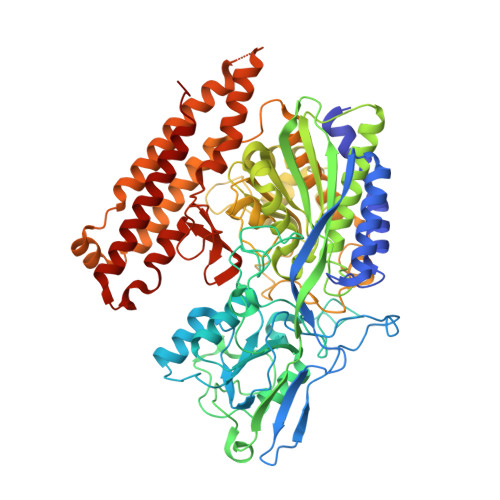
    8BO8, 8BOL, 8BOW

  • PubMed Abstract: 

    PSMA-617 is recognized as a benchmark ligand for prostate-specific membrane antigen (PSMA) owing to its broad utilization in prostate cancer (PCa) targeted radionuclide therapy. In this study, the structure-activity relationships (SAR) of PSMA-617 and two novel analogs featuring modified linkers were investigated. In compounds P17 and P18, the 2-naphthyl-l-Ala moiety was replaced with a less lipophilic 3-styryl-l-Ala moiety while the cyclohexyl ring in P18 was replaced with a phenyl group. The first ever crystal structure of the PSMA/PSMA-617 complex reported here revealed a folded conformation of the PSMA-617 linker while for the PSMA/P17 and PSMA/P18 complexes, the extended orientations of the linkers revealed linker flexibility within the PSMA cavity, a change in binding that can be exploited for the structure-guided design of PSMA-targeting agents. Despite structural differences from PSMA-617, the analogs maintained high PSMA inhibition potency, cellular binding, and internalization. In vivo biodistribution studies revealed comparable tumor uptake across all three compounds with P18 displaying higher spleen accumulation, likely due to phenyl ring lipophilicity. These SAR findings provide a strategic framework for the rational design of PSMA ligands, paving the way for the development of next-generation theranostic agents for PCa.


  • Organizational Affiliation
    • Service Unit for Radiopharmaceuticals and Preclinical Studies, German Cancer Research Center (DKFZ), Im Neuenheimer Feld 280, 69120 Heidelberg, Germany.

Macromolecule Content 

  • Total Structure Weight: 84.55 kDa 
  • Atom Count: 6,614 
  • Modeled Residue Count: 693 
  • Deposited Residue Count: 707 
  • Unique protein chains: 1

Macromolecules

Find similar proteins by:|  3D Structure
Entity ID: 1
MoleculeChains  Sequence LengthOrganismDetailsImage
Glutamate carboxypeptidase 2707Homo sapiensMutation(s): 0 
Gene Names: FOLH1FOLHNAALAD1PSMPSMAGIG27
EC: 3.4.17.21
UniProt & NIH Common Fund Data Resources
Find proteins for Q04609 (Homo sapiens)
Explore Q04609 
Go to UniProtKB:  Q04609
PHAROS:  Q04609
GTEx:  ENSG00000086205 
Entity Groups
Sequence Clusters30% Identity50% Identity70% Identity90% Identity95% Identity100% Identity
UniProt GroupQ04609
Glycosylation
Glycosylation Sites: 7Go to GlyGen: Q04609-1
Sequence Annotations
Expand
Reference Sequence

Oligosaccharides

Help  
Entity ID: 2
MoleculeChains Length2D Diagram GlycosylationD Interactions
beta-D-mannopyranose-(1-4)-2-acetamido-2-deoxy-beta-D-glucopyranose-(1-4)-2-acetamido-2-deoxy-beta-D-glucopyranose
B, F
3N-Glycosylation
Glycosylation Resources
GlyTouCan: G15407YE
GlyCosmos: G15407YE
GlyGen: G15407YE
Entity ID: 3
MoleculeChains Length2D Diagram GlycosylationD Interactions
alpha-D-mannopyranose-(1-3)-[alpha-D-mannopyranose-(1-6)]beta-D-mannopyranose-(1-4)-2-acetamido-2-deoxy-beta-D-glucopyranose-(1-4)-2-acetamido-2-deoxy-beta-D-glucopyranose
C
5N-Glycosylation
Glycosylation Resources
GlyTouCan: G22768VO
GlyCosmos: G22768VO
GlyGen: G22768VO
Entity ID: 4
MoleculeChains Length2D Diagram GlycosylationD Interactions
2-acetamido-2-deoxy-beta-D-glucopyranose-(1-4)-2-acetamido-2-deoxy-beta-D-glucopyranose
D
2N-Glycosylation
Glycosylation Resources
GlyTouCan: G42666HT
GlyCosmos: G42666HT
GlyGen: G42666HT
Entity ID: 5
MoleculeChains Length2D Diagram GlycosylationD Interactions
beta-D-mannopyranose-(1-4)-2-acetamido-2-deoxy-beta-D-glucopyranose-(1-4)-[alpha-L-fucopyranose-(1-6)]2-acetamido-2-deoxy-beta-D-glucopyranose
E
4N-Glycosylation
Glycosylation Resources
GlyTouCan: G32152BH
GlyCosmos: G32152BH
GlyGen: G32152BH

Small Molecules

Ligands 6 Unique
IDChains Name / Formula / InChI Key2D Diagram3D Interactions
QWF
(Subject of Investigation/LOI)

Query on QWF


Download:Ideal Coordinates CCD File
N [auth A](2~{S})-2-[[(2~{S})-1-oxidanyl-1-oxidanylidene-6-[[(~{E},2~{S})-5-phenyl-2-[[4-[[2-[4,7,10-tris(2-hydroxy-2-oxoethyl)-1,4,7,10-tetrazacyclododec-1-yl]ethanoylamino]methyl]cyclohexyl]carbonylamino]pent-4-enoyl]amino]hexan-2-yl]carbamoylamino]pentanedioic acid
C47 H71 N9 O16
PDXVBYVTYTYYEQ-QMBVCIHYSA-N
NAG

Query on NAG


Download:Ideal Coordinates CCD File
K [auth A],
L [auth A]
2-acetamido-2-deoxy-beta-D-glucopyranose
C8 H15 N O6
OVRNDRQMDRJTHS-FMDGEEDCSA-N
ZN

Query on ZN


Download:Ideal Coordinates CCD File
G [auth A],
H [auth A]
ZINC ION
Zn
PTFCDOFLOPIGGS-UHFFFAOYSA-N
CA

Query on CA


Download:Ideal Coordinates CCD File
I [auth A]CALCIUM ION
Ca
BHPQYMZQTOCNFJ-UHFFFAOYSA-N
CL

Query on CL


Download:Ideal Coordinates CCD File
J [auth A]CHLORIDE ION
Cl
VEXZGXHMUGYJMC-UHFFFAOYSA-M
NA

Query on NA


Download:Ideal Coordinates CCD File
M [auth A]SODIUM ION
Na
FKNQFGJONOIPTF-UHFFFAOYSA-N

Experimental Data & Validation

Experimental Data

  • Method: X-RAY DIFFRACTION
  • Resolution: 1.55 Å
  • R-Value Free:  0.226 (Depositor), 0.198 (DCC) 
  • R-Value Work:  0.202 (Depositor) 
  • R-Value Observed: 0.203 (Depositor) 
Space Group: I 2 2 2
Unit Cell:
Length ( Å )Angle ( ˚ )
a = 101.887α = 90
b = 130.286β = 90
c = 159.702γ = 90
Software Package:
Software NamePurpose
REFMACrefinement
XDSdata reduction
Aimlessdata scaling
REFMACphasing

Structure Validation

View Full Validation Report


Ligand Structure Quality Assessment 


Entry History 

& Funding Information

Deposition Data


Funding OrganizationLocationGrant Number
Not funded--

Revision History  (Full details and data files)

  • Version 1.0: 2023-11-29
    Type: Initial release
  • Version 1.1: 2024-10-16
    Changes: Structure summary
  • Version 1.2: 2025-03-26
    Changes: Database references
  • Version 1.3: 2026-03-04
    Changes: Refinement description