8BLO

Human Urea Transporter UT-A (N-Terminal Domain Model)


Experimental Data Snapshot

  • Method: ELECTRON MICROSCOPY
  • Resolution: 2.90 Å
  • Aggregation State: PARTICLE 
  • Reconstruction Method: SINGLE PARTICLE 

wwPDB Validation   3D Report Full Report


This is version 1.1 of the entry. See complete history


Literature

Structural characterization of human urea transporters UT-A and UT-B and their inhibition.

Chi, G.Dietz, L.Tang, H.Snee, M.Scacioc, A.Wang, D.Mckinley, G.Mukhopadhyay, S.M.M.Pike, A.C.W.Chalk, R.Burgess-Brown, N.A.Timmermans, J.P.van Putte, W.Robinson, C.V.Durr, K.L.

(2023) Sci Adv 9: eadg8229-eadg8229

  • DOI: https://doi.org/10.1126/sciadv.adg8229
  • Primary Citation of Related Structures:  
    8BLO, 8BLP

  • PubMed Abstract: 

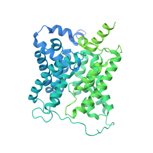
    In this study, we present the structures of human urea transporters UT-A and UT-B to characterize them at molecular level and to detail the mechanism of UT-B inhibition by its selective inhibitor, UTB inh -14. High-resolution structures of both transporters establish the structural basis for the inhibitor's selectivity to UT-B, and the identification of multiple binding sites for the inhibitor will aid with the development of drug lead molecules targeting both transporters. Our study also discovers phospholipids associating with the urea transporters by combining structural observations, native MS, and lipidomics analysis. These insights improve our understanding of urea transporter function at a molecular level and provide a blueprint for a structure-guided design of therapeutics targeting these transporters.


  • Organizational Affiliation

    Structural Genomics Consortium, Nuffield Department of Medicine, University of Oxford, Roosevelt Drive, Oxford OX3 7DQ, UK.


Macromolecules
Find similar proteins by:  (by identity cutoff)  |  3D Structure
Entity ID: 1
MoleculeChains Sequence LengthOrganismDetailsImage
Urea transporter 2A,
B [auth C],
C [auth B]
910Homo sapiensMutation(s): 0 
Gene Names: SLC14A2HUT2UT2
Membrane Entity: Yes 
UniProt & NIH Common Fund Data Resources
Find proteins for Q15849 (Homo sapiens)
Explore Q15849 
Go to UniProtKB:  Q15849
PHAROS:  Q15849
GTEx:  ENSG00000132874 
Entity Groups  
Sequence Clusters30% Identity50% Identity70% Identity90% Identity95% Identity100% Identity
UniProt GroupQ15849
Sequence Annotations
Expand
  • Reference Sequence
Small Molecules
Ligands 2 Unique
IDChains Name / Formula / InChI Key2D Diagram3D Interactions
LMN
Query on LMN

Download Ideal Coordinates CCD File 
AA [auth B]
G [auth A]
H [auth A]
I [auth A]
J [auth A]
AA [auth B],
G [auth A],
H [auth A],
I [auth A],
J [auth A],
K [auth A],
O [auth C],
P [auth C],
Q [auth C],
R [auth C],
S [auth C],
W [auth B],
X [auth B],
Y [auth B],
Z [auth B]
Lauryl Maltose Neopentyl Glycol
C47 H88 O22
MADJBYLAYPCCOO-XYPZXBMFSA-N
PLD
Query on PLD

Download Ideal Coordinates CCD File 
D [auth A]
E [auth A]
F [auth A]
L [auth C]
M [auth C]
D [auth A],
E [auth A],
F [auth A],
L [auth C],
M [auth C],
N [auth C],
T [auth B],
U [auth B],
V [auth B]
di-heneicosanoyl phosphatidyl choline
C50 H101 N O8 P
QFFSGJSMHPWZOB-QSCHNALKSA-O
Experimental Data & Validation

Experimental Data

  • Method: ELECTRON MICROSCOPY
  • Resolution: 2.90 Å
  • Aggregation State: PARTICLE 
  • Reconstruction Method: SINGLE PARTICLE 
EM Software:
TaskSoftware PackageVersion
RECONSTRUCTIONcryoSPARC

Structure Validation

View Full Validation Report


Entry History & Funding Information

Deposition Data


Funding OrganizationLocationGrant Number
Wellcome TrustUnited Kingdom106169/Z/14/Z
European Union (EU)European Union875510, 115766

Revision History  (Full details and data files)

  • Version 1.0: 2023-10-04
    Type: Initial release
  • Version 1.1: 2023-10-11
    Changes: Database references, Source and taxonomy