8BL8 | pdb_00008bl8

Human serotonin 5-HT3A receptor (apo, active/distorted conformation)


Experimental Data Snapshot

  • Method: ELECTRON MICROSCOPY
  • Resolution: 3.21 Å
  • Aggregation State: PARTICLE 
  • Reconstruction Method: SINGLE PARTICLE 

wwPDB Validation 3D Report Full Report

Validation slider image for 8BL8

This is version 1.3 of the entry. See complete history

Literature

Structural determinants for activity of the antidepressant vortioxetine at human and rodent 5-HT 3 receptors.

Lopez-Sanchez, U.Munro, L.J.Ladefoged, L.K.Pedersen, A.J.Brun, C.C.Lyngby, S.M.Baud, D.Juillan-Binard, C.Pedersen, M.G.Lummis, S.C.R.Bang-Andersen, B.Schiott, B.Chipot, C.Schoehn, G.Neyton, J.Dehez, F.Nury, H.Kristensen, A.S.

(2024) Nat Struct Mol Biol 31: 1232-1242

  • DOI: https://doi.org/10.1038/s41594-024-01282-x
  • Primary Citation Related Structures: 
    8AW2, 8AXD, 8BL8, 8BLA, 8BLB

  • PubMed Abstract: 

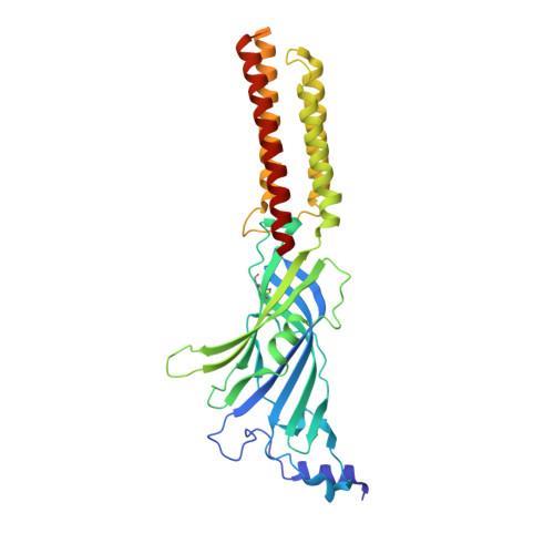
    Vortioxetine (VTX) is a recently approved antidepressant that targets a variety of serotonin receptors. Here, we investigate the drug's molecular mechanism of operation at the serotonin 5-HT 3 receptor (5-HT 3 R), which features two properties: VTX acts differently on rodent and human 5-HT 3 R, and VTX appears to suppress any subsequent response to agonists. Using a combination of cryo-EM, electrophysiology, voltage-clamp fluorometry and molecular dynamics, we show that VTX stabilizes a resting inhibited state of the mouse 5-HT 3 R and an agonist-bound-like state of human 5-HT 3 R, in line with the functional profile of the drug. We report four human 5-HT 3 R structures and show that the human receptor transmembrane domain is intrinsically fragile. We also explain the lack of recovery after VTX administration via a membrane partition mechanism.


  • Organizational Affiliation
    • University Grenoble Alpes, CNRS, CEA, IBS, Grenoble, France.

Macromolecule Content 

  • Total Structure Weight: 237.28 kDa 
  • Atom Count: 14,019 
  • Modeled Residue Count: 1,656 
  • Deposited Residue Count: 1,985 
  • Unique protein chains: 1

Macromolecules

Find similar proteins by:|  3D Structure
Entity ID: 1
MoleculeChains  Sequence LengthOrganismDetailsImage
5-hydroxytryptamine receptor 3A
A, B, C, D, E
397Homo sapiensMutation(s): 0 
Gene Names: HTR3A5HT3RHTR3
Membrane Entity: Yes 
UniProt & NIH Common Fund Data Resources
Find proteins for P46098 (Homo sapiens)
Explore P46098 
Go to UniProtKB:  P46098
PHAROS:  P46098
GTEx:  ENSG00000166736 
Entity Groups
Sequence Clusters30% Identity50% Identity70% Identity90% Identity95% Identity100% Identity
UniProt GroupP46098
Glycosylation
Glycosylation Sites: 3Go to GlyGen: P46098-1
Sequence Annotations
Expand
Reference Sequence

Oligosaccharides

Help  
Entity ID: 2
MoleculeChains Length2D Diagram GlycosylationD Interactions
2-acetamido-2-deoxy-beta-D-glucopyranose-(1-4)-2-acetamido-2-deoxy-beta-D-glucopyranose
F, G, H, I, J
F, G, H, I, J, K, L, M, N, O
2N-Glycosylation
Glycosylation Resources
GlyTouCan: G42666HT
GlyCosmos: G42666HT
GlyGen: G42666HT

Experimental Data & Validation

Experimental Data

  • Method: ELECTRON MICROSCOPY
  • Resolution: 3.21 Å
  • Aggregation State: PARTICLE 
  • Reconstruction Method: SINGLE PARTICLE 
EM Software:
TaskSoftware PackageVersion
MODEL REFINEMENTPHENIX

Structure Validation

View Full Validation Report


Entry History 

& Funding Information

Deposition Data


Funding OrganizationLocationGrant Number
European Research Council (ERC)European Union637733

Revision History  (Full details and data files)

  • Version 1.0: 2024-05-15
    Type: Initial release
  • Version 1.1: 2024-08-28
    Changes: Data collection, Database references
  • Version 1.2: 2024-11-20
    Changes: Data collection, Structure summary
  • Version 1.3: 2025-07-02
    Changes: Data collection