Characterization of a unique repression system present in arbitrium phages of the SPbeta family.
Brady, A., Cabello-Yeves, E., Gallego Del Sol, F., Chmielowska, C., Mancheno-Bonillo, J., Zamora-Caballero, S., Omer, S.B., Torres-Puente, M., Eldar, A., Quiles-Puchalt, N., Marina, A., Penades, J.R.(2023) Cell Host Microbe 31: 2023-2037.e8
- PubMed: 38035880 Search on PubMed
- DOI: https://doi.org/10.1016/j.chom.2023.11.003
- Primary Citation Related Structures:
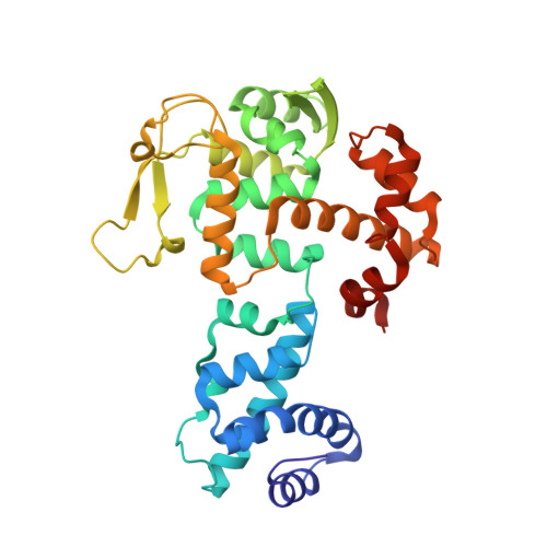
8BJ6, 8BJV, 8BPZ - PubMed Abstract:
Arbitrium-coding phages use peptides to communicate and coordinate the decision between lysis and lysogeny. However, the mechanism by which these phages establish lysogeny remains unknown. Here, focusing on the SPbeta phage family's model phages phi3T and SPβ, we report that a six-gene operon called the "SPbeta phages repressor operon" (sro) expresses not one but two master repressors, SroE and SroF, the latter of which folds like a classical phage integrase. To promote lysogeny, these repressors bind to multiple sites in the phage genome. SroD serves as an auxiliary repressor that, with SroEF, forms the repression module necessary for lysogeny establishment and maintenance. Additionally, the proteins SroABC within the operon are proposed to constitute the transducer module, connecting the arbitrium communication system to the activity of the repression module. Overall, this research sheds light on the intricate and specialized repression system employed by arbitrium SPβ-like phages in making lysis-lysogeny decisions.
- Centre for Bacterial Resistance Biology, Imperial College London, London SW7 2AZ, UK; Institute of Infection, Immunity and Inflammation, University of Glasgow, Glasgow G12 8TA, UK.
Organizational Affiliation: