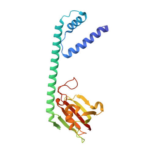
Legionella pneumophila macrophage infectivity potentiator protein appendage domains modulate protein dynamics and inhibitor binding.
Wiedemann, C., Whittaker, J.J., Perez Carrillo, V.H., Goretzki, B., Dajka, M., Tebbe, F., Harder, J.M., Krajczy, P.R., Joseph, B., Hausch, F., Guskov, A., Hellmich, U.A.(2023) Int J Biol Macromol 252: 126366-126366
- PubMed: 37633566 Search on PubMed
- DOI: https://doi.org/10.1016/j.ijbiomac.2023.126366
- Primary Citation Related Structures:
8BJC, 8BJD, 8BJE, 8BK4, 8BK5, 8BK6 - PubMed Abstract:
Macrophage infectivity potentiator (MIP) proteins are widespread in human pathogens including Legionella pneumophila, the causative agent of Legionnaires' disease and protozoans such as Trypanosoma cruzi. All MIP proteins contain a FKBP (FK506 binding protein)-like prolyl-cis/trans-isomerase domain that hence presents an attractive drug target. Some MIPs such as the Legionella pneumophila protein (LpMIP) have additional appendage domains of mostly unknown function. In full-length, homodimeric LpMIP, the N-terminal dimerization domain is linked to the FKBP-like domain via a long, free-standing stalk helix. Combining X-ray crystallography, NMR and EPR spectroscopy and SAXS, we elucidated the importance of the stalk helix for protein dynamics and inhibitor binding to the FKBP-like domain and bidirectional crosstalk between the different protein regions. The first comparison of a microbial MIP and a human FKBP in complex with the same synthetic inhibitor was made possible by high-resolution structures of LpMIP with a [4.3.1]-aza-bicyclic sulfonamide and provides a basis for designing pathogen-selective inhibitors. Through stereospecific methylation, the affinity of inhibitors to L. pneumophila and T. cruzi MIP was greatly improved. The resulting X-ray inhibitor-complex structures of LpMIP and TcMIP at 1.49 and 1.34 Å, respectively, provide a starting point for developing potent inhibitors against MIPs from multiple pathogenic microorganisms.
- Faculty of Chemistry and Earth Sciences, Institute of Organic Chemistry and Macromolecular Chemistry, Friedrich Schiller University Jena, Jena, Germany.
Organizational Affiliation: