8BJ9

X-ray structure of the CeuE Homologue from Parageobacillus thermoglucosidasius - 5LICAM complex.


Experimental Data Snapshot

  • Method: X-RAY DIFFRACTION
  • Resolution: 2.07 Å
  • R-Value Free: 0.266 
  • R-Value Work: 0.210 

wwPDB Validation   3D Report Full Report


Ligand Structure Quality Assessment 


This is version 1.3 of the entry. See complete history


Literature

Thermostable homologues of the periplasmic siderophore-binding protein CeuE from Geobacillus stearothermophilus and Parageobacillus thermoglucosidasius.

Blagova, E.V.Miller, A.H.Bennett, M.Booth, R.L.Dodson, E.J.Duhme-Klair, A.K.Wilson, K.S.

(2023) Acta Crystallogr D Struct Biol 79: 694-705

  • DOI: https://doi.org/10.1107/S2059798323004473
  • Primary Citation of Related Structures:  
    8B7X, 8BAW, 8BAX, 8BF6, 8BJ9, 8BNW

  • PubMed Abstract: 

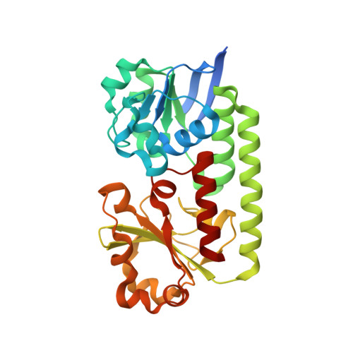
    Siderophore-binding proteins from two thermophilic bacteria, Geobacillus stearothermophilus and Parageobacillus thermoglucosidasius, were identified from a search of sequence databases, cloned and overexpressed. They are homologues of the well characterized protein CjCeuE from Campylobacter jejuni. The iron-binding histidine and tyrosine residues are conserved in both thermophiles. Crystal structures were determined of the apo proteins and of their complexes with iron(III)-azotochelin and its analogue iron(III)-5-LICAM. The thermostability of both homologues was shown to be about 20°C higher than that of CjCeuE. Similarly, the tolerance of the homologues to the organic solvent dimethylformamide (DMF) was enhanced, as reflected by the respective binding constants for these ligands measured in aqueous buffer at pH 7.5 in the absence and presence of 10% and 20% DMF. Consequently, these thermophilic homologues offer advantages in the development of artificial metalloenzymes using the CeuE family.


  • Organizational Affiliation

    Structural Biology Laboratory, Department of Chemistry, University of York, Heslington, York YO10 5DD, United Kingdom.


Macromolecules
Find similar proteins by:  (by identity cutoff)  |  3D Structure
Entity ID: 1
MoleculeChains Sequence LengthOrganismDetailsImage
ABC transporter297Parageobacillus thermoglucosidasiusMutation(s): 0 
Gene Names: BAA00_15370
Entity Groups  
Sequence Clusters30% Identity50% Identity70% Identity90% Identity95% Identity100% Identity
Sequence Annotations
Expand
  • Reference Sequence
Small Molecules
Ligands 4 Unique
IDChains Name / Formula / InChI Key2D Diagram3D Interactions
5LC (Subject of Investigation/LOI)
Query on 5LC

Download Ideal Coordinates CCD File 
C [auth A]N,N'-pentane-1,5-diylbis(2,3-dihydroxybenzamide)
C19 H22 N2 O6
BRWLZTCRFJVZDD-UHFFFAOYSA-N
SO4
Query on SO4

Download Ideal Coordinates CCD File 
H [auth A]SULFATE ION
O4 S
QAOWNCQODCNURD-UHFFFAOYSA-L
NI
Query on NI

Download Ideal Coordinates CCD File 
D [auth A],
E [auth A],
F [auth A],
G [auth A]
NICKEL (II) ION
Ni
VEQPNABPJHWNSG-UHFFFAOYSA-N
FE (Subject of Investigation/LOI)
Query on FE

Download Ideal Coordinates CCD File 
B [auth A]FE (III) ION
Fe
VTLYFUHAOXGGBS-UHFFFAOYSA-N
Experimental Data & Validation

Experimental Data

  • Method: X-RAY DIFFRACTION
  • Resolution: 2.07 Å
  • R-Value Free: 0.266 
  • R-Value Work: 0.210 
  • Space Group: C 2 2 21
Unit Cell:
Length ( Å )Angle ( ˚ )
a = 36.063α = 90
b = 117.392β = 90
c = 141.712γ = 90
Software Package:
Software NamePurpose
REFMACrefinement
REFMACrefinement
DIALSdata reduction
Aimlessdata scaling
MOLREPphasing

Structure Validation

View Full Validation Report


Ligand Structure Quality Assessment 


Entry History & Funding Information

Deposition Data


Funding OrganizationLocationGrant Number
UK Research and Innovation (UKRI)United KingdomEP/T007338/1

Revision History  (Full details and data files)

  • Version 1.0: 2023-07-12
    Type: Initial release
  • Version 1.1: 2023-07-19
    Changes: Database references
  • Version 1.2: 2023-08-09
    Changes: Data collection, Database references
  • Version 1.3: 2024-02-07
    Changes: Data collection, Refinement description