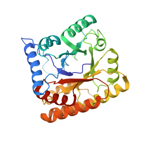
Crystal structure of FAD-independent methylene-tetrahydrofolate reductase from Mycobacterium hassiacum.
Gehl, M., Demmer, U., Ermler, U., Shima, S.(2023) Proteins 91: 1329-1340
- PubMed: 37119125 Search on PubMed
- DOI: https://doi.org/10.1002/prot.26504
- Primary Citation Related Structures:
8BGR - PubMed Abstract:
FAD-independent methylene-tetrahydrofolate (methylene-H 4 F) reductase (Mfr), recently identified in mycobacteria, catalyzes the reduction of methylene-H 4 F to methyl-H 4 F with NADH as hydride donor by a ternary complex mechanism. This biochemical reaction corresponds to that of the ubiquitous FAD-dependent methylene-H 4 F reductase (MTHFR), although the latter uses a ping-pong mechanism with the prosthetic group as intermediate hydride carrier. Comparative genomics and genetic analyses indicated that Mfr is indispensable for the growth of Mycobacterium tuberculosis, which lacks the MTHFR encoding gene. Therefore, Mfr appears to be an excellent target for the design of antimycobacterial drugs. Here, we report the heterologous production, enzymological characterization, and the crystal structure of Mfr from the thermophilic mycobacterium Mycobacterium hassiacum (hMfr), which shows 78% sequence identity to Mfr from M. tuberculosis. Although hMfr and MTHFR have minor sequence identity and different catalytic mechanisms, their structures are highly similar, thus suggesting a divergent evolution of Mfr and MTHFR from a common ancestor. Most of the important active site residues of MTHFR are conserved and equivalently positioned in the tertiary structure of hMfr. The Glu9Gln variant of hMfr exhibits a drastic reduction of the catalytic activity, which supports the predicted function of the glutamate residue as proton donor in both hMfr and MTHFR. Thus, highly similar binding modes for the C 1 -carriers and the reducing agents in hMfr and MTHFR are assumed.
- Max Planck Institute for Terrestrial Microbiology, Marburg, Germany.
Organizational Affiliation: