8BB6 | pdb_00008bb6

Crystal structure of Arabidopsis thaliana sucrose transporter SUC1


Experimental Data Snapshot

  • Method: X-RAY DIFFRACTION
  • Resolution: 2.68 Å
  • R-Value Free: 
    0.293 (Depositor), 0.294 (DCC) 
  • R-Value Work: 
    0.270 (Depositor), 0.272 (DCC) 
  • R-Value Observed: 
    0.271 (Depositor) 

Starting Model: in silico
View more details

wwPDB Validation 3D Report Full Report

Validation slider image for 8BB6

This is version 1.3 of the entry. See complete history

Literature

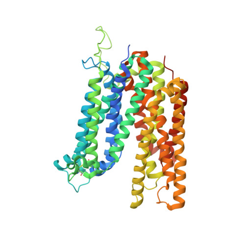
Structure and sucrose binding mechanism of the plant SUC1 sucrose transporter.

Bavnhoj, L.Driller, J.H.Zuzic, L.Stange, A.D.Schiott, B.Pedersen, B.P.

(2023) Nat Plants 9: 938-950

  • DOI: https://doi.org/10.1038/s41477-023-01421-0
  • Primary Citation Related Structures: 
    8BB6

  • PubMed Abstract: 

    Sucrose import from photosynthetic tissues into the phloem is mediated by transporters from the low-affinity sucrose transporter family (SUC/SUT family). Furthermore, sucrose redistribution to other tissues is driven by phloem sap movement, the product of high turgor pressure created by this import activity. Additionally, sink organs such as fruits, cereals and seeds that accumulate high concentrations of sugar also depend on this active transport of sucrose. Here we present the structure of the sucrose-proton symporter, Arabidopsis thaliana SUC1, in an outward open conformation at 2.7 Å resolution, together with molecular dynamics simulations and biochemical characterization. We identify the key acidic residue required for proton-driven sucrose uptake and describe how protonation and sucrose binding are strongly coupled. Sucrose binding is a two-step process, with initial recognition mediated by the glucosyl moiety binding directly to the key acidic residue in a stringent pH-dependent manner. Our results explain how low-affinity sucrose transport is achieved in plants, and pinpoint a range of SUC binders that help define selectivity. Our data demonstrate a new mode for proton-driven symport with links to cation-driven symport and provide a broad model for general low-affinity transport in highly enriched substrate environments.


  • Organizational Affiliation
    • Department of Molecular Biology and Genetics, Aarhus University, Aarhus, Denmark.

Macromolecule Content 

  • Total Structure Weight: 110.97 kDa 
  • Atom Count: 7,220 
  • Modeled Residue Count: 952 
  • Deposited Residue Count: 1,038 
  • Unique protein chains: 1

Macromolecules

Find similar proteins by:|  3D Structure
Entity ID: 1
MoleculeChains  Sequence LengthOrganismDetailsImage
Sucrose transport protein SUC1
A, B
519Arabidopsis thalianaMutation(s): 0 
Gene Names: SUC1At1g71880F17M19.3
Membrane Entity: Yes 
UniProt
Find proteins for Q39232 (Arabidopsis thaliana)
Explore Q39232 
Go to UniProtKB:  Q39232
Entity Groups
Sequence Clusters30% Identity50% Identity70% Identity90% Identity95% Identity100% Identity
UniProt GroupQ39232
Sequence Annotations
Expand
Reference Sequence

Experimental Data & Validation

Experimental Data

  • Method: X-RAY DIFFRACTION
  • Resolution: 2.68 Å
  • R-Value Free:  0.293 (Depositor), 0.294 (DCC) 
  • R-Value Work:  0.270 (Depositor), 0.272 (DCC) 
  • R-Value Observed: 0.271 (Depositor) 
Space Group: P 1
Unit Cell:
Length ( Å )Angle ( ˚ )
a = 58.776α = 89.96
b = 65.622β = 101.74
c = 82.813γ = 94.72
Software Package:
Software NamePurpose
PHENIXrefinement
xia2data scaling
PHASERphasing
PDB_EXTRACTdata extraction
DIALSdata reduction

Structure Validation

View Full Validation Report


Entry History 

& Funding Information

Deposition Data


Funding OrganizationLocationGrant Number
European Research Council (ERC)European Union101000936

Revision History  (Full details and data files)

  • Version 1.0: 2023-05-31
    Type: Initial release
  • Version 1.1: 2023-06-28
    Changes: Database references
  • Version 1.2: 2024-05-01
    Changes: Data collection, Refinement description
  • Version 1.3: 2024-11-06
    Changes: Structure summary