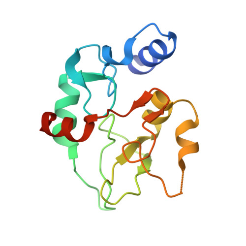
The MECP2-TRD domain interacts with the DNMT3A-ADD domain at the H3-tail binding site.
Kunert, S., Linhard, V., Weirich, S., Choudalakis, M., Osswald, F., Kramer, L., Kohler, A.R., Brohm, A., Wollenhaupt, J., Schwalbe, H., Jeltsch, A.(2023) Protein Sci 32: e4542-e4542
- PubMed: 36519786 Search on PubMedSearch on PubMed Central
- DOI: https://doi.org/10.1002/pro.4542
- Primary Citation Related Structures:
8BA5 - PubMed Abstract:
The DNMT3A DNA methyltransferase and MECP2 methylation reader are highly expressed in neurons. Both proteins interact via their DNMT3A-ADD and MECP2-TRD domains, and the MECP2 interaction regulates the activity and subnuclear localization of DNMT3A. Here, we mapped the interface of both domains using peptide SPOT array binding, protein pull-down, equilibrium peptide binding assays, and structural analyses. The region D529-D531 on the surface of the ADD domain was identified as interaction point with the TRD domain. This includes important residues of the histone H3 N-terminal tail binding site to the ADD domain, explaining why TRD and H3 binding to the ADD domain is competitive. On the TRD domain, residues 214-228 containing K219 and K223 were found to be essential for the ADD interaction. This part represents a folded patch within the otherwise largely disordered TRD domain. A crystal structure analysis of ADD revealed that the identified H3/TDR lysine binding pocket is occupied by an arginine residue from a crystallographic neighbor in the ADD apoprotein structure. Finally, we show that mutations in the interface of ADD and TRD domains disrupt the cellular interaction of both proteins in NIH3T3 cells. In summary, our data show that the H3 peptide binding cleft of the ADD domain also mediates the interaction with the MECP2-TRD domain suggesting that this binding site may have a broader role also in the interaction of DNMT3A with other proteins leading to complex regulation options by competitive and PTM specific binding.
- Institute of Biochemistry and Technical Biochemistry, University of Stuttgart, Stuttgart, Germany.
Organizational Affiliation: