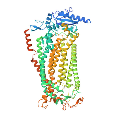
Structural basis for the activation of the lipid scramblase TMEM16F.
Arndt, M., Alvadia, C., Straub, M.S., Clerico Mosina, V., Paulino, C., Dutzler, R.(2022) Nat Commun 13: 6692-6692
- PubMed: 36335104 Search on PubMedSearch on PubMed Central
- DOI: https://doi.org/10.1038/s41467-022-34497-x
- Primary Citation Related Structures:
8B8G, 8B8J, 8B8K, 8B8M, 8B8Q, 8BC0, 8BC1 - PubMed Abstract:
TMEM16F, a member of the conserved TMEM16 family, plays a central role in the initiation of blood coagulation and the fusion of trophoblasts. The protein mediates passive ion and lipid transport in response to an increase in intracellular Ca 2+ . However, the mechanism of how the protein facilitates both processes has remained elusive. Here we investigate the basis for TMEM16F activation. In a screen of residues lining the proposed site of conduction, we identify mutants with strongly activating phenotype. Structures of these mutants determined herein by cryo-electron microscopy show major rearrangements leading to the exposure of hydrophilic patches to the membrane, whose distortion facilitates lipid diffusion. The concomitant opening of a pore promotes ion conduction in the same protein conformation. Our work has revealed a mechanism that is distinct for this branch of the family and that will aid the development of a specific pharmacology for a promising drug target.
- Department of Biochemistry University of Zurich, Winterthurer Str. 190, CH-8057, Zurich, Switzerland.
Organizational Affiliation: