8B8H | pdb_00008b8h

Structure of DCS-resistant variant D322N of alanine racemase from M. tuberculosis in complex with DCS


Experimental Data Snapshot

  • Method: X-RAY DIFFRACTION
  • Resolution: 1.78 Å
  • R-Value Free: 
    0.223 (Depositor), 0.223 (DCC) 
  • R-Value Work: 
    0.199 (Depositor), 0.199 (DCC) 
  • R-Value Observed: 
    0.200 (Depositor) 

Starting Model: experimental
View more details

wwPDB Validation 3D Report Full Report

Validation slider image for 8B8H

Ligand Structure Quality Assessment 


This is version 1.2 of the entry. See complete history

Literature

Structure of the d-Cycloserine-Resistant Variant D322N of Alanine Racemase from Mycobacterium tuberculosis .

de Chiara, C.Prosser, G.A.Ogrodowicz, R.de Carvalho, L.P.S.

(2023) ACS Bio Med Chem Au 3: 233-239

  • DOI: https://doi.org/10.1021/acsbiomedchemau.2c00074
  • Primary Citation Related Structures: 
    8AHW, 8B8H

  • PubMed Abstract: 

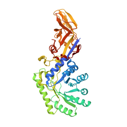
    Alanine racemase (Alr) is a pyridoxal 5'-phosphate-dependent enzyme that catalyzes the racemization of l-alanine to d-alanine. Alr is one of the two targets of the broad-spectrum antibiotic d-cycloserine (DCS), a structural analogue of d-alanine. Despite being an essential component of regimens used to treat multi- and extensively drug-resistant tuberculosis for almost seven decades, resistance to DCS has not been observed in patients. We previously demonstrated that DCS evades resistance due to an ultralow rate of emergence of mutations. Yet, we identified a single polymorphism (converting Asp322 to Asn) in the alr gene, which arose in 8 out of 11 independent variants identified and that confers resistance. Here, we present the crystal structure of the Alr variant D322N in both the free and DCS-inactivated forms and the characterization of its DCS inactivation mechanism by UV-visible and fluorescence spectroscopy. Comparison of these results with those obtained with wild-type Alr reveals the structural basis of the 240-fold reduced inhibition observed in Alr D322N.


  • Organizational Affiliation
    • Mycobacterial Metabolism and Antibiotic Research Laboratory, The Francis Crick Institute, London, NW1 1AT, United Kingdom.

Macromolecule Content 

  • Total Structure Weight: 83.49 kDa 
  • Atom Count: 5,927 
  • Modeled Residue Count: 742 
  • Deposited Residue Count: 774 
  • Unique protein chains: 1

Macromolecules

Find similar proteins by:|  3D Structure
Entity ID: 1
MoleculeChains  Sequence LengthOrganismDetailsImage
Alanine racemase
A, B
387Mycobacterium tuberculosis H37RvMutation(s): 1 
Gene Names: alrRv3423cMTCY78.06
EC: 5.1.1.1
UniProt
Find proteins for P9WQA9 (Mycobacterium tuberculosis (strain ATCC 25618 / H37Rv))
Explore P9WQA9 
Go to UniProtKB:  P9WQA9
Entity Groups
Sequence Clusters30% Identity50% Identity70% Identity90% Identity95% Identity100% Identity
UniProt GroupP9WQA9
Sequence Annotations
Expand
Reference Sequence

Small Molecules

Ligands 6 Unique
IDChains Name / Formula / InChI Key2D Diagram3D Interactions
L7N
(Subject of Investigation/LOI)

Query on L7N


Download:Ideal Coordinates CCD File
D [auth A],
H [auth B]
(~{E})-[2-methyl-3-oxidanyl-5-(phosphonooxymethyl)pyridin-4-yl]methylidene-[(4~{R})-3-oxidanylidene-1,2-oxazolidin-4-yl]azanium
C11 H15 N3 O7 P
PWALIMMCDFLLIJ-RZTFRGLUSA-O
OJQ
(Subject of Investigation/LOI)

Query on OJQ


Download:Ideal Coordinates CCD File
C [auth A][2-methyl-3-oxidanyl-5-(phosphonooxymethyl)pyridin-4-yl]methyl-(3-oxidanyl-1,2-oxazol-4-yl)azanium
C11 H15 N3 O7 P
PXWFNGNWQUPGPJ-UHFFFAOYSA-O
GOL

Query on GOL


Download:Ideal Coordinates CCD File
G [auth A]GLYCEROL
C3 H8 O3
PEDCQBHIVMGVHV-UHFFFAOYSA-N
EDO

Query on EDO


Download:Ideal Coordinates CCD File
J [auth B],
K [auth B],
L [auth B]
1,2-ETHANEDIOL
C2 H6 O2
LYCAIKOWRPUZTN-UHFFFAOYSA-N
CA

Query on CA


Download:Ideal Coordinates CCD File
E [auth A]CALCIUM ION
Ca
BHPQYMZQTOCNFJ-UHFFFAOYSA-N
CL

Query on CL


Download:Ideal Coordinates CCD File
F [auth A],
I [auth B]
CHLORIDE ION
Cl
VEXZGXHMUGYJMC-UHFFFAOYSA-M

Experimental Data & Validation

Experimental Data

  • Method: X-RAY DIFFRACTION
  • Resolution: 1.78 Å
  • R-Value Free:  0.223 (Depositor), 0.223 (DCC) 
  • R-Value Work:  0.199 (Depositor), 0.199 (DCC) 
  • R-Value Observed: 0.200 (Depositor) 
Space Group: P 41 21 2
Unit Cell:
Length ( Å )Angle ( ˚ )
a = 163.94α = 90
b = 163.94β = 90
c = 57.11γ = 90
Software Package:
Software NamePurpose
DIALSdata reduction
xia2data scaling
PHASERphasing
PHENIXrefinement

Structure Validation

View Full Validation Report


Ligand Structure Quality Assessment 


Entry History 

& Funding Information

Deposition Data


Funding OrganizationLocationGrant Number
The Francis Crick InstituteUnited KingdomSee list below
Cancer Research UKUnited KingdomFC001060
Medical Research Council (MRC, United Kingdom)United KingdomFC001060
Wellcome TrustUnited KingdomFC001060
Wellcome TrustUnited Kingdom104785/B/14/Z

Revision History  (Full details and data files)

  • Version 1.0: 2023-04-05
    Type: Initial release
  • Version 1.1: 2023-07-05
    Changes: Database references
  • Version 1.2: 2024-02-07
    Changes: Data collection, Refinement description