


The structure of Gan1D W433A in complex with galactose-6P
Snyder, J., Lansky, S., Zehavi, A., Shoham, Y., Shoham, G.To be published.
Experimental Data Snapshot
Starting Model: experimental
View more details
Entity ID: 1 | |||||
|---|---|---|---|---|---|
| Molecule | Chains | Sequence Length | Organism | Details | Image |
| Putative 6-phospho-beta-galactobiosidase | 478 | Geobacillus stearothermophilus | Mutation(s): 1 Gene Names: gan1D EC: 3.2.1.85 | 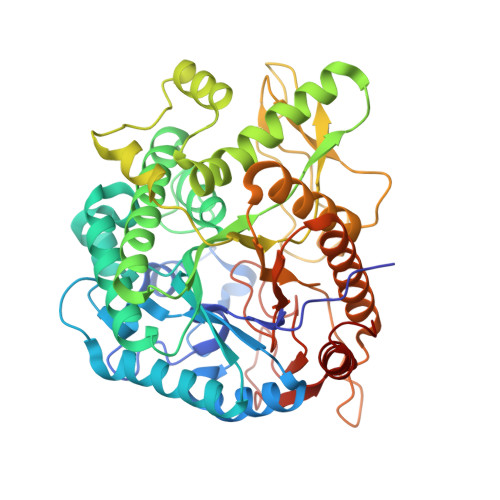 | |
UniProt | |||||
Entity Groups | |||||
| Sequence Clusters | 30% Identity50% Identity70% Identity90% Identity95% Identity100% Identity | ||||
| UniProt Group | W8QF82 | ||||
Sequence AnnotationsExpand | |||||
Reference Sequence | |||||
| Ligands 3 Unique | |||||
|---|---|---|---|---|---|
| ID | Chains | Name / Formula / InChI Key | 2D Diagram | 3D Interactions | |
| BGP (Subject of Investigation/LOI) Download:Ideal Coordinates CCD File | F [auth A] | 6-O-phosphono-beta-D-galactopyranose C6 H13 O9 P NBSCHQHZLSJFNQ-FPRJBGLDSA-N |  | ||
| GOL Download:Ideal Coordinates CCD File | G [auth A] | GLYCEROL C3 H8 O3 PEDCQBHIVMGVHV-UHFFFAOYSA-N |  | ||
| IMD Download:Ideal Coordinates CCD File | C [auth A], D [auth A], E [auth A] | IMIDAZOLE C3 H5 N2 RAXXELZNTBOGNW-UHFFFAOYSA-O |  | ||
| Length ( Å ) | Angle ( ˚ ) |
|---|---|
| a = 107.5 | α = 90 |
| b = 68.6 | β = 99.62 |
| c = 152.29 | γ = 90 |
| Software Name | Purpose |
|---|---|
| Aimless | data scaling |
| PHENIX | refinement |
| PDB_EXTRACT | data extraction |
| XDS | data reduction |
| PHENIX | phasing |
| Funding Organization | Location | Grant Number |
|---|---|---|
| Not funded | Israel | -- |


