


Crystal structure of Quinonoid dihydropteridine reductase from Leishmania donovani
Robinson, D.A., Fairlamb, A.H.To be published.
Experimental Data Snapshot
Starting Model: experimental
View more details
wwPDB Validation 3D Report Full Report

Entity ID: 1 | |||||
|---|---|---|---|---|---|
| Molecule | Chains | Sequence Length | Organism | Details | Image |
| Quinonoid dihydropteridine reductase | 250 | Leishmania donovani | Mutation(s): 0 Gene Names: CGC21_27355, LdCL_340051200, LdCL_340051500, LDHU3_34.6510 EC: 1.5.1.34 | 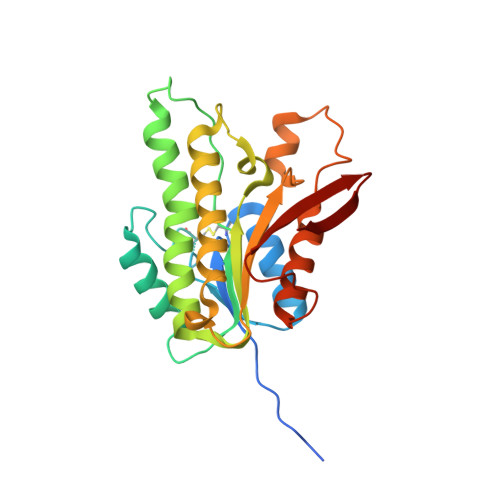 | |
UniProt | |||||
Entity Groups | |||||
| Sequence Clusters | 30% Identity50% Identity70% Identity90% Identity95% Identity100% Identity | ||||
| UniProt Group | A0A3S5H7Y1 | ||||
Sequence AnnotationsExpand | |||||
Reference Sequence | |||||
| Length ( Å ) | Angle ( ˚ ) |
|---|---|
| a = 66.762 | α = 90 |
| b = 66.762 | β = 90 |
| c = 242.92 | γ = 120 |
| Software Name | Purpose |
|---|---|
| Aimless | data scaling |
| REFMAC | refinement |
| PDB_EXTRACT | data extraction |
| XDS | data reduction |
| MOLREP | phasing |
| Funding Organization | Location | Grant Number |
|---|---|---|
| Not funded | -- |


