Structural and time-resolved mechanistic investigations of protein hydrolysis by the acidic proline-specific endoprotease from Aspergillus niger.
Pijning, T., Vujicic-Zagar, A., van der Laan, J.M., de Jong, R.M., Ramirez-Palacios, C., Vente, A., Edens, L., Dijkstra, B.W.(2024) Protein Sci 33: e4856-e4856
- PubMed: 38059672 Search on PubMedSearch on PubMed Central
- DOI: https://doi.org/10.1002/pro.4856
- Primary Citation Related Structures:
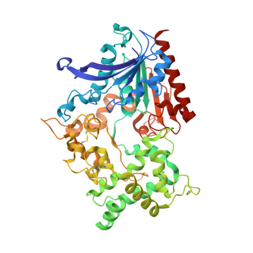
8B57, 8BBX - PubMed Abstract:
Proline-specific endoproteases have been successfully used in, for example, the in-situ degradation of gluten, the hydrolysis of bitter peptides, the reduction of haze during beer production, and the generation of peptides for mass spectroscopy and proteomics applications. Here we present the crystal structure of the extracellular proline-specific endoprotease from Aspergillus niger (AnPEP), a member of the S28 peptidase family with rarely observed true proline-specific endoprotease activity. Family S28 proteases have a conventional Ser-Asp-His catalytic triad, but their oxyanion-stabilizing hole shows a glutamic acid, an amino acid not previously observed in this role. Since these enzymes have an acidic pH optimum, the presence of a glutamic acid in the oxyanion hole may confine their activity to an acidic pH. Yet, considering the presence of the conventional catalytic triad, it is remarkable that the A. niger enzyme remains active down to pH 1.5. The determination of the primary cleavage site of cytochrome c along with molecular dynamics-assisted docking studies indicate that the active site pocket of AnPEP can accommodate a reverse turn of approximately 12 amino acids with proline at the S1 specificity pocket. Comparison with the structures of two S28-proline-specific exopeptidases reveals not only a more spacious active site cavity but also the absence of any putative binding sites for amino- and carboxyl-terminal residues as observed in the exopeptidases, explaining AnPEP's observed endoprotease activity.
- Biomolecular X-ray Crystallography, Groningen Biomolecular Sciences and Biotechnology Institute (GBB), University of Groningen, Groningen, The Netherlands.
Organizational Affiliation: