8B3B

Structure of metacyclic VSG (mVSG) 531 from Trypanosoma brucei


Experimental Data Snapshot

  • Method: X-RAY DIFFRACTION
  • Resolution: 1.95 Å
  • R-Value Free: 0.238 
  • R-Value Work: 0.200 
  • R-Value Observed: 0.202 

wwPDB Validation   3D Report Full Report


Ligand Structure Quality Assessment 


This is version 1.2 of the entry. See complete history


Literature

Structural similarities between the metacyclic and bloodstream form variant surface glycoproteins of the African trypanosome.

Chandra, M.Dakovic, S.Foti, K.Zeelen, J.P.van Straaten, M.Aresta-Branco, F.Tihon, E.Lubbehusen, N.Ruppert, T.Glover, L.Papavasiliou, F.N.Stebbins, C.E.

(2023) PLoS Negl Trop Dis 17: e0011093-e0011093

  • DOI: https://doi.org/10.1371/journal.pntd.0011093
  • Primary Citation of Related Structures:  
    8B3B, 8B3E, 8B3W

  • PubMed Abstract: 

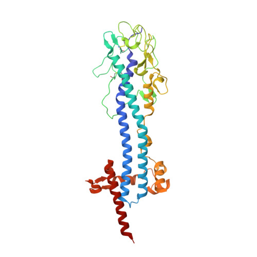
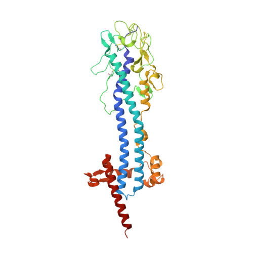
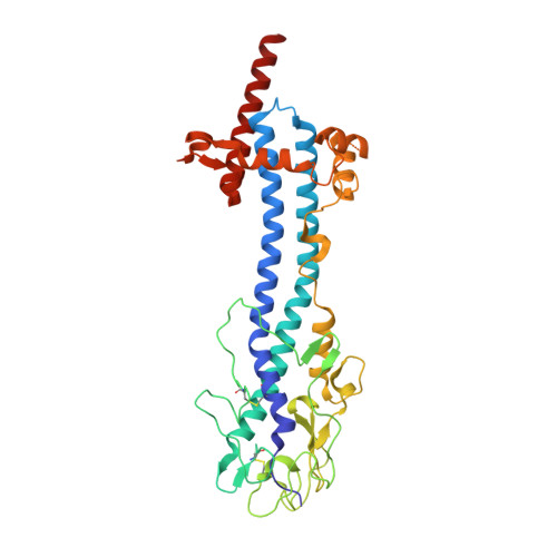
    During infection of mammalian hosts, African trypanosomes thwart immunity using antigenic variation of the dense Variant Surface Glycoprotein (VSG) coat, accessing a large repertoire of several thousand genes and pseudogenes, and switching to antigenically distinct copies. The parasite is transferred to mammalian hosts by the tsetse fly. In the salivary glands of the fly, the pathogen adopts the metacyclic form and expresses a limited repertoire of VSG genes specific to that developmental stage. It has remained unknown whether the metacyclic VSGs possess distinct properties associated with this particular and discrete phase of the parasite life cycle. We present here three novel metacyclic form VSG N-terminal domain crystal structures (mVSG397, mVSG531, and mVSG1954) and show that they mirror closely in architecture, oligomerization, and surface diversity the known classes of bloodstream form VSGs. These data suggest that the mVSGs are unlikely to be a specialized subclass of VSG proteins, and thus could be poor candidates as the major components of prophylactic vaccines against trypanosomiasis.


  • Organizational Affiliation

    Division of Structural Biology of Infection and Immunity, German Cancer Research Center, Heidelberg, Germany.


Macromolecules
Find similar proteins by:  (by identity cutoff)  |  3D Structure
Entity ID: 1
MoleculeChains Sequence LengthOrganismDetailsImage
Variant surface glycoprotein 531A,
B [auth C]
371Trypanosoma bruceiMutation(s): 0 
UniProt
Find proteins for M4SYA9 (Trypanosoma brucei)
Explore M4SYA9 
Go to UniProtKB:  M4SYA9
Entity Groups  
Sequence Clusters30% Identity50% Identity70% Identity90% Identity95% Identity100% Identity
UniProt GroupM4SYA9
Sequence Annotations
Expand
  • Reference Sequence
Find similar proteins by:  (by identity cutoff)  |  3D Structure
Entity ID: 2
MoleculeChains Sequence LengthOrganismDetailsImage
Variant surface glycoprotein 531C [auth D],
E [auth G],
H
372Trypanosoma bruceiMutation(s): 0 
UniProt
Find proteins for M4SYA9 (Trypanosoma brucei)
Explore M4SYA9 
Go to UniProtKB:  M4SYA9
Entity Groups  
Sequence Clusters30% Identity50% Identity70% Identity90% Identity95% Identity100% Identity
UniProt GroupM4SYA9
Sequence Annotations
Expand
  • Reference Sequence
Find similar proteins by:  (by identity cutoff)  |  3D Structure
Entity ID: 3
MoleculeChains Sequence LengthOrganismDetailsImage
Variant surface glycoprotein 531D [auth B],
F [auth E],
G [auth F]
370Trypanosoma bruceiMutation(s): 0 
UniProt
Find proteins for M4SYA9 (Trypanosoma brucei)
Explore M4SYA9 
Go to UniProtKB:  M4SYA9
Entity Groups  
Sequence Clusters30% Identity50% Identity70% Identity90% Identity95% Identity100% Identity
UniProt GroupM4SYA9
Sequence Annotations
Expand
  • Reference Sequence
Oligosaccharides

Help

Entity ID: 4
MoleculeChains Length2D Diagram Glycosylation3D Interactions
alpha-D-mannopyranose-(1-2)-alpha-D-mannopyranose-(1-3)-[alpha-D-mannopyranose-(1-6)]beta-D-mannopyranose-(1-4)-2-acetamido-2-deoxy-beta-D-glucopyranose-(1-4)-2-acetamido-2-deoxy-beta-D-glucopyranose
I, J
6N-Glycosylation
Glycosylation Resources
GlyTouCan:  G56014GC
GlyCosmos:  G56014GC
GlyGen:  G56014GC
Small Molecules
Ligands 3 Unique
IDChains Name / Formula / InChI Key2D Diagram3D Interactions
NAG
Query on NAG

Download Ideal Coordinates CCD File 
DA [auth E]
EA [auth E]
JA [auth F]
K [auth C]
KA [auth F]
DA [auth E],
EA [auth E],
JA [auth F],
K [auth C],
KA [auth F],
L [auth C],
OA [auth H],
PA [auth H],
R [auth D],
S [auth D],
X [auth B],
Y [auth B]
2-acetamido-2-deoxy-beta-D-glucopyranose
C8 H15 N O6
OVRNDRQMDRJTHS-FMDGEEDCSA-N
MAN
Query on MAN

Download Ideal Coordinates CCD File 
AA [auth B]
BA [auth B]
CA [auth B]
FA [auth E]
GA [auth E]
AA [auth B],
BA [auth B],
CA [auth B],
FA [auth E],
GA [auth E],
HA [auth E],
IA [auth E],
MA [auth F],
N [auth C],
NA [auth F],
O [auth C],
P [auth C],
Q [auth C],
RA [auth H],
SA [auth H],
TA [auth H],
U [auth D],
V [auth D],
W [auth D]
alpha-D-mannopyranose
C6 H12 O6
WQZGKKKJIJFFOK-PQMKYFCFSA-N
BMA
Query on BMA

Download Ideal Coordinates CCD File 
LA [auth F],
M [auth C],
QA [auth H],
T [auth D],
Z [auth B]
beta-D-mannopyranose
C6 H12 O6
WQZGKKKJIJFFOK-RWOPYEJCSA-N
Experimental Data & Validation

Experimental Data

  • Method: X-RAY DIFFRACTION
  • Resolution: 1.95 Å
  • R-Value Free: 0.238 
  • R-Value Work: 0.200 
  • R-Value Observed: 0.202 
  • Space Group: P 1 21 1
Unit Cell:
Length ( Å )Angle ( ˚ )
a = 50.107α = 90
b = 170.885β = 90.71
c = 163.652γ = 90
Software Package:
Software NamePurpose
PHENIXrefinement

Structure Validation

View Full Validation Report


Ligand Structure Quality Assessment 


Entry History & Funding Information

Deposition Data


Funding OrganizationLocationGrant Number
Other governmentGermany--

Revision History  (Full details and data files)

  • Version 1.0: 2023-01-25
    Type: Initial release
  • Version 1.1: 2023-04-19
    Changes: Database references
  • Version 1.2: 2024-02-07
    Changes: Data collection, Refinement description