8B0U | pdb_00008b0u

Structure of the CalpL/T10 complex


Experimental Data Snapshot

  • Method: X-RAY DIFFRACTION
  • Resolution: 3.29 Å
  • R-Value Free: 
    0.230 (Depositor), 0.230 (DCC) 
  • R-Value Work: 
    0.195 (Depositor), 0.200 (DCC) 
  • R-Value Observed: 
    0.197 (Depositor) 

Starting Model: experimental
View more details

wwPDB Validation   3D Report Full Report


This is version 1.3 of the entry. See complete history


Literature

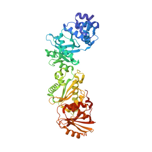
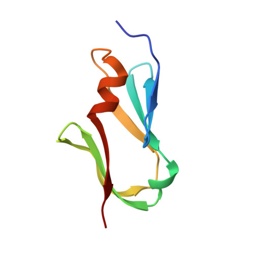
Antiviral signalling by a cyclic nucleotide activated CRISPR protease.

Rouillon, C.Schneberger, N.Chi, H.Blumenstock, K.Da Vela, S.Ackermann, K.Moecking, J.Peter, M.F.Boenigk, W.Seifert, R.Bode, B.E.Schmid-Burgk, J.L.Svergun, D.Geyer, M.White, M.F.Hagelueken, G.

(2023) Nature 614: 168-174

  • DOI: https://doi.org/10.1038/s41586-022-05571-7
  • Primary Citation of Related Structures:  
    7QDA, 8B0R, 8B0U

  • PubMed Abstract: 

    CRISPR defence systems such as the well-known DNA-targeting Cas9 and the RNA-targeting type III systems are widespread in prokaryotes 1,2 . The latter orchestrates a complex antiviral response that is initiated through the synthesis of cyclic oligoadenylates after recognition of foreign RNA 3-5 . Among the large set of proteins that are linked to type III systems and predicted to bind cyclic oligoadenylates 6,7 , a CRISPR-associated Lon protease (CalpL) stood out to us. CalpL contains a sensor domain of the SAVED family 7 fused to a Lon protease effector domain. However, the mode of action of this effector is unknown. Here we report the structure and function of CalpL and show that this soluble protein forms a stable tripartite complex with two other proteins, CalpT and CalpS, that are encoded on the same operon. After activation by cyclic tetra-adenylate (cA 4 ), CalpL oligomerizes and specifically cleaves the MazF homologue CalpT, which releases the extracytoplasmic function σ factor CalpS from the complex. Our data provide a direct connection between CRISPR-based detection of foreign nucleic acids and transcriptional regulation. Furthermore, the presence of a SAVED domain that binds cyclic tetra-adenylate in a CRISPR effector reveals a link to the cyclic-oligonucleotide-based antiphage signalling system.


  • Organizational Affiliation
    • Institute of Structural Biology, University of Bonn, Bonn, Germany. back2crispr@gmail.com.

Macromolecules
Find similar proteins by:  (by identity cutoff)  |  3D Structure
Entity ID: 1
MoleculeChains Sequence LengthOrganismDetailsImage
SAVED domain-containing proteinA,
C [auth B]
496SulfurihydrogenibiumMutation(s): 0 
Gene Names: SYO3AOP1_0656
Entity Groups  
Sequence Clusters30% Identity50% Identity70% Identity90% Identity95% Identity100% Identity
Sequence Annotations
Expand
  • Reference Sequence
Find similar proteins by:  (by identity cutoff)  |  3D Structure
Entity ID: 2
MoleculeChains Sequence LengthOrganismDetailsImage
CalpT10B [auth D],
D [auth C]
76SulfurihydrogenibiumMutation(s): 0 
Gene Names: SYO3AOP1_0655
Entity Groups  
Sequence Clusters30% Identity50% Identity70% Identity90% Identity95% Identity100% Identity
Sequence Annotations
Expand
  • Reference Sequence
Small Molecules
Ligands 2 Unique
IDChains Name / Formula / InChI Key2D Diagram3D Interactions
SO4
Query on SO4

Download Ideal Coordinates CCD File 
E [auth A]
F [auth A]
G [auth A]
H [auth A]
K [auth B]
E [auth A],
F [auth A],
G [auth A],
H [auth A],
K [auth B],
L [auth B],
M [auth B]
SULFATE ION
O4 S
QAOWNCQODCNURD-UHFFFAOYSA-L
GOL
Query on GOL

Download Ideal Coordinates CCD File 
I [auth D],
J [auth D]
GLYCEROL
C3 H8 O3
PEDCQBHIVMGVHV-UHFFFAOYSA-N
Experimental Data & Validation

Experimental Data

  • Method: X-RAY DIFFRACTION
  • Resolution: 3.29 Å
  • R-Value Free:  0.230 (Depositor), 0.230 (DCC) 
  • R-Value Work:  0.195 (Depositor), 0.200 (DCC) 
  • R-Value Observed: 0.197 (Depositor) 
Space Group: P 1 21 1
Unit Cell:
Length ( Å )Angle ( ˚ )
a = 89.754α = 90
b = 96.598β = 98.19
c = 131.71γ = 90
Software Package:
Software NamePurpose
PHASERphasing

Structure Validation

View Full Validation Report


Entry History & Funding Information

Deposition Data


Funding OrganizationLocationGrant Number
German Research Foundation (DFG)GermanyHA 6805/6-1

Revision History  (Full details and data files)

  • Version 1.0: 2022-11-16
    Type: Initial release
  • Version 1.1: 2023-01-11
    Changes: Database references, Structure summary
  • Version 1.2: 2023-02-08
    Changes: Database references
  • Version 1.3: 2024-02-07
    Changes: Data collection, Refinement description