8B08 | pdb_00008b08

TRYPTOPHAN SYNTHASE - Cryo-trapping by the spitrobot crystal plunger after 30 sec


Experimental Data Snapshot

  • Method: X-RAY DIFFRACTION
  • Resolution: 2.50 Å
  • R-Value Free: 
    0.242 (Depositor), 0.242 (DCC) 
  • R-Value Work: 
    0.197 (Depositor), 0.197 (DCC) 
  • R-Value Observed: 
    0.199 (Depositor) 

Starting Model: experimental
View more details

wwPDB Validation 3D Report Full Report

Validation slider image for 8B08

Ligand Structure Quality Assessment 


This is version 1.2 of the entry. See complete history

Literature

Millisecond cryo-trapping by the spitrobot crystal plunger simplifies time-resolved crystallography.

Mehrabi, P.Sung, S.von Stetten, D.Prester, A.Hatton, C.E.Kleine-Dopke, S.Berkes, A.Gore, G.Leimkohl, J.P.Schikora, H.Kollewe, M.Rohde, H.Wilmanns, M.Tellkamp, F.Schulz, E.C.

(2023) Nat Commun 14: 2365-2365

  • DOI: https://doi.org/10.1038/s41467-023-37834-w
  • Primary Citation Related Structures: 
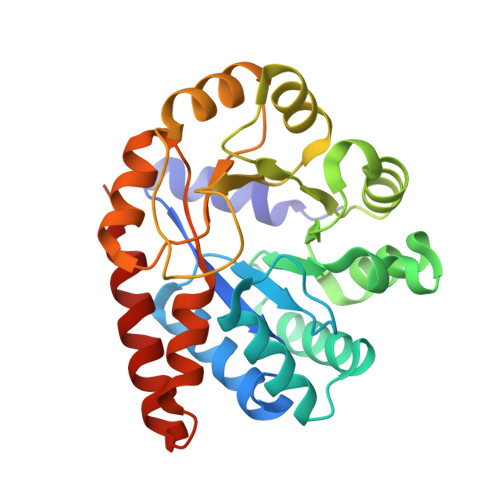
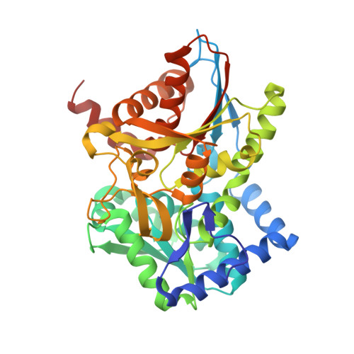
    8AW8, 8AW9, 8AWB, 8AWC, 8AWD, 8AWE, 8AWF, 8AWS, 8AWU, 8AWV, 8AWX, 8AWY, 8B03, 8B05, 8B06, 8B08, 8B2O, 8B2V, 8B2W, 8B3M

  • PubMed Abstract: 

    We introduce the spitrobot, a protein crystal plunger, enabling reaction quenching via cryo-trapping with a time-resolution in the millisecond range. Protein crystals are mounted on canonical micromeshes on an electropneumatic piston, where the crystals are kept in a humidity and temperature-controlled environment, then reactions are initiated via the liquid application method (LAMA) and plunging into liquid nitrogen is initiated after an electronically set delay time to cryo-trap intermediate states. High-magnification images are automatically recorded before and after droplet deposition, prior to plunging. The SPINE-standard sample holder is directly plunged into a storage puck, enabling compatibility with high-throughput infrastructure. Here we demonstrate binding of glucose and 2,3-butanediol in microcrystals of xylose isomerase, and of avibactam and ampicillin in microcrystals of the extended spectrum beta-lactamase CTX-M-14. We also trap reaction intermediates and conformational changes in macroscopic crystals of tryptophan synthase to demonstrate that the spitrobot enables insight into catalytic events.


  • Organizational Affiliation
    • Institute for Nanostructure and Solid-State Physics, Universität Hamburg, Hamburg, Germany. pedram.mehrabi@uni-hamburg.de.

Macromolecule Content 

  • Total Structure Weight: 72.47 kDa 
  • Atom Count: 5,173 
  • Modeled Residue Count: 663 
  • Deposited Residue Count: 666 
  • Unique protein chains: 2

Macromolecules

Find similar proteins by:|  3D Structure
Entity ID: 1
MoleculeChains  Sequence LengthOrganismDetailsImage
Tryptophan synthase alpha chain269Salmonella enterica subsp. enterica serovar TyphimuriumMutation(s): 0 
Gene Names: trpASTM1727
EC: 4.2.1.20
UniProt
Find proteins for P00929 (Salmonella typhimurium (strain LT2 / SGSC1412 / ATCC 700720))
Explore P00929 
Go to UniProtKB:  P00929
Entity Groups
Sequence Clusters30% Identity50% Identity70% Identity90% Identity95% Identity100% Identity
UniProt GroupP00929
Sequence Annotations
Expand
Reference Sequence
Find similar proteins by:|  3D Structure
Entity ID: 2
MoleculeChains  Sequence LengthOrganismDetailsImage
Tryptophan synthase beta chain397Salmonella enterica subsp. enterica serovar TyphimuriumMutation(s): 0 
Gene Names: trpBSTM1726
EC: 4.2.1.20
UniProt
Find proteins for P0A2K1 (Salmonella typhimurium (strain LT2 / SGSC1412 / ATCC 700720))
Explore P0A2K1 
Go to UniProtKB:  P0A2K1
Entity Groups
Sequence Clusters30% Identity50% Identity70% Identity90% Identity95% Identity100% Identity
UniProt GroupP0A2K1
Sequence Annotations
Expand
Reference Sequence

Small Molecules

Ligands 4 Unique
IDChains Name / Formula / InChI Key2D Diagram3D Interactions
PLS
(Subject of Investigation/LOI)

Query on PLS


Download:Ideal Coordinates CCD File
E [auth B][3-HYDROXY-2-METHYL-5-PHOSPHONOOXYMETHYL-PYRIDIN-4-YLMETHYL]-SERINE
C11 H17 N2 O8 P
ODVKKQWXKRZJLG-VIFPVBQESA-N
G3P
(Subject of Investigation/LOI)

Query on G3P


Download:Ideal Coordinates CCD File
C [auth A]SN-GLYCEROL-3-PHOSPHATE
C3 H9 O6 P
AWUCVROLDVIAJX-GSVOUGTGSA-N
CS

Query on CS


Download:Ideal Coordinates CCD File
D [auth B]CESIUM ION
Cs
NCMHKCKGHRPLCM-UHFFFAOYSA-N
CL

Query on CL


Download:Ideal Coordinates CCD File
F [auth B],
G [auth B]
CHLORIDE ION
Cl
VEXZGXHMUGYJMC-UHFFFAOYSA-M

Experimental Data & Validation

Experimental Data

  • Method: X-RAY DIFFRACTION
  • Resolution: 2.50 Å
  • R-Value Free:  0.242 (Depositor), 0.242 (DCC) 
  • R-Value Work:  0.197 (Depositor), 0.197 (DCC) 
  • R-Value Observed: 0.199 (Depositor) 
Space Group: C 1 2 1
Unit Cell:
Length ( Å )Angle ( ˚ )
a = 182.482α = 90
b = 60.093β = 94.75
c = 67.341γ = 90
Software Package:
Software NamePurpose
XDSdata reduction
Aimlessdata scaling
PHENIXrefinement
PDB_EXTRACTdata extraction
PHASERphasing

Structure Validation

View Full Validation Report


Ligand Structure Quality Assessment 


Entry History 

& Funding Information

Deposition Data


Funding OrganizationLocationGrant Number
H2020 Marie Curie Actions of the European CommissionEuropean Union664726

Revision History  (Full details and data files)

  • Version 1.0: 2023-05-03
    Type: Initial release
  • Version 1.1: 2023-05-24
    Changes: Database references
  • Version 1.2: 2024-02-07
    Changes: Data collection, Refinement description