8AYJ | pdb_00008ayj

Crystal structure of D-amino acid aminotransferase from Aminobacterium colombiens complexed with 3-aminooxypropionic acid


Experimental Data Snapshot

  • Method: X-RAY DIFFRACTION
  • Resolution: 1.75 Å
  • R-Value Free: 
    0.239 (Depositor), 0.245 (DCC) 
  • R-Value Work: 
    0.201 (Depositor), 0.209 (DCC) 
  • R-Value Observed: 
    0.203 (Depositor) 

Starting Model: experimental
View more details

wwPDB Validation 3D Report Full Report

Validation slider image for 8AYJ

Ligand Structure Quality Assessment 


This is version 1.2 of the entry. See complete history

Literature

In search for structural targets for engineering d-amino acid transaminase: modulation of pH optimum and substrate specificity.

Shilova, S.A.Matyuta, I.O.Khrenova, M.G.Nikolaeva, A.Y.Klyachko, N.L.Minyaev, M.E.Khomutov, A.R.Boyko, K.M.Popov, V.O.Bezsudnova, E.Y.

(2023) Biochem J 480: 1267-1284

  • DOI: https://doi.org/10.1042/BCJ20230233
  • Primary Citation Related Structures: 
    8AYJ, 8ONJ, 8ONL, 8ONN

  • PubMed Abstract: 

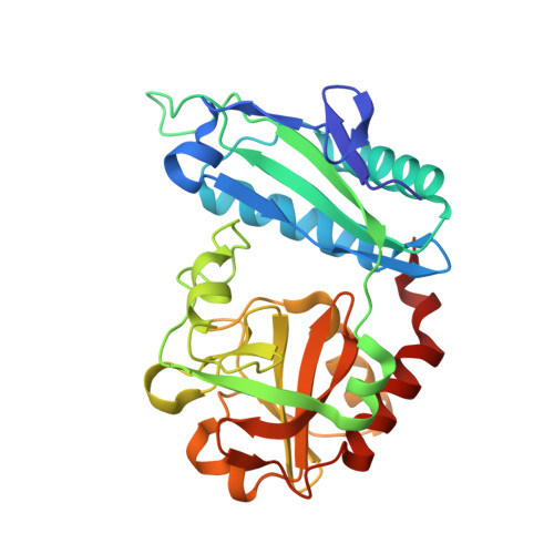
    The development of biocatalysts requires reorganization of the enzyme's active site to facilitate the productive binding of the target substrate and improve turnover number at desired conditions. Pyridoxal-5'-phosphate (PLP) - dependent transaminases are highly efficient biocatalysts for asymmetric amination of ketones and keto acids. However, transaminases, being stereoselective enzymes, have a narrow substrate specificity due to the ordered structure of the active site and work only in neutral-alkaline media. Here, we investigated the d-amino acid transaminase from Aminobacterium colombiense, with the active site organized differently from that of the canonical d-amino acid transaminase from Bacillus sp. YM-1. Using a combination of site-directed mutagenesis, kinetic analysis, molecular modeling, and structural analysis we determined the active site residues responsible for substrate binding, substrate differentiation, thermostability of a functional dimer, and affecting the pH optimum. We demonstrated that the high specificity toward d-glutamate/α-ketoglutarate is due to the interactions of a γ-carboxylate group with K237 residue, while binding of other substrates stems from the effectiveness of their accommodation in the active site optimized for d-glutamate/α-ketoglutarate binding. Furthermore, we showed that the K237A substitution shifts the catalytic activity optimum to acidic pH. Our findings are useful for achieving target substrate specificity and demonstrate the potential for developing and optimizing transaminases for various applications.


  • Organizational Affiliation
    • Bach Institute of Biochemistry, Research Centre of Biotechnology of the Russian Academy of Sciences, Moscow, Russia.

Macromolecule Content 

  • Total Structure Weight: 63.85 kDa 
  • Atom Count: 4,928 
  • Modeled Residue Count: 552 
  • Deposited Residue Count: 554 
  • Unique protein chains: 1

Macromolecules

Find similar proteins by:|  3D Structure
Entity ID: 1
MoleculeChains  Sequence LengthOrganismDetailsImage
Aminotransferase class IV
A, B
277Aminobacterium colombienseMutation(s): 0 
Gene Names: Amico_1844
UniProt
Find proteins for D5EHC5 (Aminobacterium colombiense (strain DSM 12261 / ALA-1))
Explore D5EHC5 
Go to UniProtKB:  D5EHC5
Entity Groups
Sequence Clusters30% Identity50% Identity70% Identity90% Identity95% Identity100% Identity
UniProt GroupD5EHC5
Sequence Annotations
Expand
Reference Sequence

Small Molecules

Ligands 3 Unique
IDChains Name / Formula / InChI Key2D Diagram3D Interactions
OCF

Query on OCF


Download:Ideal Coordinates CCD File
C [auth A],
E [auth A],
G [auth B],
I [auth B]
3-[(~{E})-[2-methyl-3-oxidanyl-5-(phosphonooxymethyl)pyridin-4-yl]methylideneamino]oxypropanoic acid
C11 H15 N2 O8 P
WFKOVQOIGDESOJ-WLRTZDKTSA-N
PLP

Query on PLP


Download:Ideal Coordinates CCD File
F [auth A],
H [auth B]
PYRIDOXAL-5'-PHOSPHATE
C8 H10 N O6 P
NGVDGCNFYWLIFO-UHFFFAOYSA-N
EDO

Query on EDO


Download:Ideal Coordinates CCD File
D [auth A]1,2-ETHANEDIOL
C2 H6 O2
LYCAIKOWRPUZTN-UHFFFAOYSA-N

Experimental Data & Validation

Experimental Data

  • Method: X-RAY DIFFRACTION
  • Resolution: 1.75 Å
  • R-Value Free:  0.239 (Depositor), 0.245 (DCC) 
  • R-Value Work:  0.201 (Depositor), 0.209 (DCC) 
  • R-Value Observed: 0.203 (Depositor) 
Space Group: P 1 21 1
Unit Cell:
Length ( Å )Angle ( ˚ )
a = 50.573α = 90
b = 58.434β = 94.11
c = 96.722γ = 90
Software Package:
Software NamePurpose
REFMACrefinement
Aimlessdata scaling
PDB_EXTRACTdata extraction
XDSdata reduction
REFMACphasing

Structure Validation

View Full Validation Report


Ligand Structure Quality Assessment 


Entry History 

& Funding Information

Deposition Data


Funding OrganizationLocationGrant Number
Russian Science FoundationRussian Federation19-14-00164

Revision History  (Full details and data files)

  • Version 1.0: 2022-11-16
    Type: Initial release
  • Version 1.1: 2023-08-30
    Changes: Data collection, Database references, Refinement description
  • Version 1.2: 2024-01-31
    Changes: Refinement description