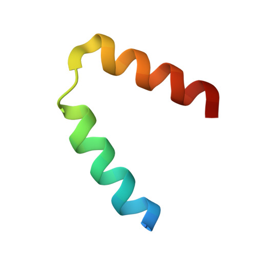
The C-terminus of CFAP410 forms a tetrameric helical bundle that is essential for its localization to the basal body.
Stadler, A., De Liz, L.V., Gabriel, H.B., Alonso-Gil, S., Crickley, R., Korbula, K., Zagrovic, B., Vaughan, S., Sunter, J.D., Dong, G.(2024) Open Biol 14: 240128-240128
- PubMed: 39255848 Search on PubMed
- DOI: https://doi.org/10.1098/rsob.240128
- Primary Citation Related Structures:
8AXO, 8AXR, 8R9T - PubMed Abstract:
Cilia are antenna-like organelles protruding from the surface of many cell types in the human body. Defects in ciliary structure or function often lead to diseases that are collectively called ciliopathies. Cilia and flagella-associated protein 410 (CFAP410) localizes at the basal body of cilia/flagella and plays essential roles in ciliogenesis, neuronal development and DNA damage repair. It remains unknown how its specific basal body location is achieved. Multiple single amino acid mutations in CFAP410 have been identified in patients with various ciliopathies. One of the mutations, L224P, is located in the C-terminal domain (CTD) of human CFAP410 and causes severe spondylometaphyseal dysplasia, axial (SMDAX). However, the molecular mechanism for how the mutation causes the disorder remains unclear. Here, we report our structural studies on the CTD of CFAP410 from three distantly related organisms, Homo sapiens, Trypanosoma brucei and Chlamydomonas reinhardtii . The crystal structures reveal that the three proteins all adopt the same conformation as a tetrameric helical bundle. Our work further demonstrates that the tetrameric assembly of the CTD is essential for the correct localization of CFAP410 in T. brucei , as the L224P mutation that disassembles the tetramer disrupts its basal body localization. Taken together, our studies reveal that the basal body localization of CFAP410 is controlled by the CTD and provide a mechanistic explanation for how the mutation L224P in CFAP410 causes ciliopathies in humans.
- Max Perutz Labs, Vienna Biocenter, Medical University of Vienna, Vienna 1030, Austria.
Organizational Affiliation: