8AR7 | pdb_00008ar7

Bovine glutamate dehydrogenase in ternary complex with the allosteric activators ADP and leucine


Experimental Data Snapshot

  • Method: X-RAY DIFFRACTION
  • Resolution: 2.45 Å
  • R-Value Free: 
    0.214 (Depositor), 0.210 (DCC) 
  • R-Value Work: 
    0.192 (Depositor), 0.187 (DCC) 
  • R-Value Observed: 
    0.193 (Depositor) 

Starting Model: experimental
View more details

wwPDB Validation 3D Report Full Report

Validation slider image for 8AR7

Ligand Structure Quality Assessment 


This is version 1.2 of the entry. See complete history

Literature

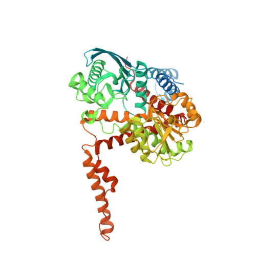
Structural Basis for the Binding of Allosteric Activators Leucine and ADP to Mammalian Glutamate Dehydrogenase.

Aleshin, V.A.Bunik, V.I.Bruch, E.M.Bellinzoni, M.

(2022) Int J Mol Sci 23

  • DOI: https://doi.org/10.3390/ijms231911306
  • Primary Citation Related Structures: 
    8AR7, 8AR8

  • PubMed Abstract: 

    Glutamate dehydrogenase (GDH) plays a key role in the metabolism of glutamate, an important compound at a cross-road of carbon and nitrogen metabolism and a relevant neurotransmitter. Despite being one of the first discovered allosteric enzymes, GDH still poses challenges for structural characterization of its allosteric sites. Only the structures with ADP, and at low (3.5 Å) resolution, are available for mammalian GDH complexes with allosteric activators. Here, we aim at deciphering a structural basis for the GDH allosteric activation using bovine GDH as a model. For the first time, we report a mammalian GDH structure in a ternary complex with the activators leucine and ADP, co-crystallized with potassium ion, resolved to 2.45 Å. An improved 2.4-angstrom resolution of the GDH complex with ADP is also presented. The ternary complex with leucine and ADP differs from the binary complex with ADP by the conformation of GDH C-terminus, involved in the leucine binding and subunit interactions. The potassium site, identified in this work, may mediate interactions between the leucine and ADP binding sites. Our data provide novel insights into the mechanisms of GDH activation by leucine and ADP, linked to the enzyme regulation by (de)acetylation.


  • Organizational Affiliation
    • Department of Biokinetics, A. N. Belozersky Institute of Physicochemical Biology, Lomonosov Moscow State University, 119234 Moscow, Russia.

Macromolecule Content 

  • Total Structure Weight: 373.88 kDa 
  • Atom Count: 23,655 
  • Modeled Residue Count: 2,982 
  • Deposited Residue Count: 3,366 
  • Unique protein chains: 1

Macromolecules

Find similar proteins by:|  3D Structure
Entity ID: 1
MoleculeChains  Sequence LengthOrganismDetailsImage
Glutamate dehydrogenase (NAD(P)(+))
A, B, C, D, E
A, B, C, D, E, F
561Bos taurusMutation(s): 0 
EC: 1.4.1.3
UniProt
Find proteins for A0A140T871 (Bos taurus)
Explore A0A140T871 
Go to UniProtKB:  A0A140T871
Entity Groups
Sequence Clusters30% Identity50% Identity70% Identity90% Identity95% Identity100% Identity
UniProt GroupA0A140T871
Sequence Annotations
Expand
Reference Sequence

Small Molecules

Ligands 3 Unique
IDChains Name / Formula / InChI Key2D Diagram3D Interactions
ADP
(Subject of Investigation/LOI)

Query on ADP


Download:Ideal Coordinates CCD File
G [auth A]
K [auth B]
N [auth C]
R [auth D]
U [auth E]
G [auth A],
K [auth B],
N [auth C],
R [auth D],
U [auth E],
W [auth F]
ADENOSINE-5'-DIPHOSPHATE
C10 H15 N5 O10 P2
XTWYTFMLZFPYCI-KQYNXXCUSA-N
LEU
(Subject of Investigation/LOI)

Query on LEU


Download:Ideal Coordinates CCD File
I [auth A]
J [auth A]
M [auth B]
P [auth C]
Q [auth D]
I [auth A],
J [auth A],
M [auth B],
P [auth C],
Q [auth D],
T [auth E]
LEUCINE
C6 H13 N O2
ROHFNLRQFUQHCH-YFKPBYRVSA-N
K

Query on K


Download:Ideal Coordinates CCD File
H [auth A]
L [auth B]
O [auth C]
S [auth D]
V [auth E]
H [auth A],
L [auth B],
O [auth C],
S [auth D],
V [auth E],
X [auth F]
POTASSIUM ION
K
NPYPAHLBTDXSSS-UHFFFAOYSA-N

Experimental Data & Validation

Experimental Data

  • Method: X-RAY DIFFRACTION
  • Resolution: 2.45 Å
  • R-Value Free:  0.214 (Depositor), 0.210 (DCC) 
  • R-Value Work:  0.192 (Depositor), 0.187 (DCC) 
  • R-Value Observed: 0.193 (Depositor) 
Space Group: P 1 21 1
Unit Cell:
Length ( Å )Angle ( ˚ )
a = 90.877α = 90
b = 178.705β = 104
c = 123.884γ = 90
Software Package:
Software NamePurpose
BUSTERrefinement
autoPROCdata reduction
STARANISOdata scaling
PHASERphasing

Structure Validation

View Full Validation Report


Ligand Structure Quality Assessment 


Entry History 

& Funding Information

Deposition Data


Funding OrganizationLocationGrant Number
Pasteur InstituteFrance--

Revision History  (Full details and data files)

  • Version 1.0: 2022-10-05
    Type: Initial release
  • Version 1.1: 2022-10-26
    Changes: Database references
  • Version 1.2: 2024-01-31
    Changes: Data collection, Refinement description