8AP9

rotor of the Trypanosoma brucei mitochondrial ATP synthase dimer


Experimental Data Snapshot

  • Method: ELECTRON MICROSCOPY
  • Resolution: 3.70 Å
  • Aggregation State: PARTICLE 
  • Reconstruction Method: SINGLE PARTICLE 

wwPDB Validation   3D Report Full Report


This is version 1.0 of the entry. See complete history


Literature

An ancestral interaction module promotes oligomerization in divergent mitochondrial ATP synthases.

Gahura, O.Muhleip, A.Hierro-Yap, C.Panicucci, B.Jain, M.Hollaus, D.Slapnickova, M.Zikova, A.Amunts, A.

(2022) Nat Commun 13: 5989-5989

  • DOI: https://doi.org/10.1038/s41467-022-33588-z
  • Primary Citation of Related Structures:  
    8AP6, 8AP7, 8AP8, 8AP9, 8APA, 8APB, 8APC, 8APD, 8APE, 8APF, 8APG, 8APH, 8APJ, 8APK

  • PubMed Abstract: 

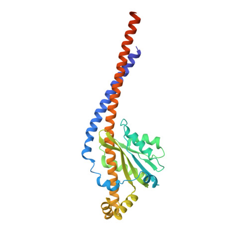
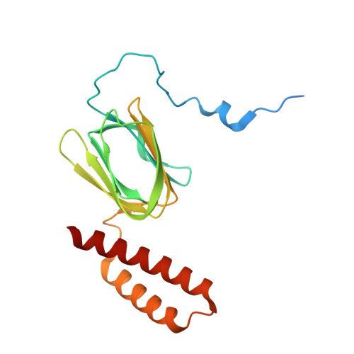
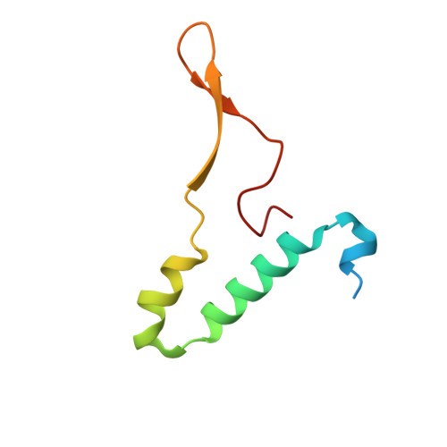
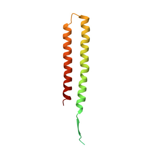
    Mitochondrial ATP synthase forms stable dimers arranged into oligomeric assemblies that generate the inner-membrane curvature essential for efficient energy conversion. Here, we report cryo-EM structures of the intact ATP synthase dimer from Trypanosoma brucei in ten different rotational states. The model consists of 25 subunits, including nine lineage-specific, as well as 36 lipids. The rotary mechanism is influenced by the divergent peripheral stalk, conferring a greater conformational flexibility. Proton transfer in the lumenal half-channel occurs via a chain of five ordered water molecules. The dimerization interface is formed by subunit-g that is critical for interactions but not for the catalytic activity. Although overall dimer architecture varies among eukaryotes, we find that subunit-g together with subunit-e form an ancestral oligomerization motif, which is shared between the trypanosomal and mammalian lineages. Therefore, our data defines the subunit-g/e module as a structural component determining ATP synthase oligomeric assemblies.


  • Organizational Affiliation

    Institute of Parasitology, Biology Centre, Czech Academy of Sciences, 37005, České Budějovice, Czech Republic.


Macromolecules
Find similar proteins by:  (by identity cutoff)  |  3D Structure
Entity ID: 1
MoleculeChains Sequence LengthOrganismDetailsImage
ATP synthase gamma subunitA [auth G]305Trypanosoma brucei bruceiMutation(s): 0 
EC: 3.6.3.14
Membrane Entity: Yes 
UniProt
Find proteins for A0A161CFW5 (Trypanosoma brucei brucei)
Explore A0A161CFW5 
Go to UniProtKB:  A0A161CFW5
Entity Groups  
Sequence Clusters30% Identity50% Identity70% Identity90% Identity95% Identity100% Identity
UniProt GroupA0A161CFW5
Sequence Annotations
Expand
  • Reference Sequence
Find similar proteins by:  (by identity cutoff)  |  3D Structure
Entity ID: 2
MoleculeChains Sequence LengthOrganismDetailsImage
ATP synthase, epsilon chain, putativeB [auth H]182Trypanosoma brucei bruceiMutation(s): 0 
EC: 3.6.3
Membrane Entity: Yes 
UniProt
Find proteins for Q586H1 (Trypanosoma brucei brucei (strain 927/4 GUTat10.1))
Explore Q586H1 
Go to UniProtKB:  Q586H1
Entity Groups  
Sequence Clusters30% Identity50% Identity70% Identity90% Identity95% Identity100% Identity
UniProt GroupQ586H1
Sequence Annotations
Expand
  • Reference Sequence
Find similar proteins by:  (by identity cutoff)  |  3D Structure
Entity ID: 3
MoleculeChains Sequence LengthOrganismDetailsImage
ATP synthase subunit epsilon, mitochondrialC [auth I]75Trypanosoma brucei bruceiMutation(s): 0 
Membrane Entity: Yes 
UniProt
Find proteins for P0DPG3 (Trypanosoma brucei brucei)
Explore P0DPG3 
Go to UniProtKB:  P0DPG3
Entity Groups  
Sequence Clusters30% Identity50% Identity70% Identity90% Identity95% Identity100% Identity
UniProt GroupP0DPG3
Sequence Annotations
Expand
  • Reference Sequence
Find similar proteins by:  (by identity cutoff)  |  3D Structure
Entity ID: 4
MoleculeChains Sequence LengthOrganismDetailsImage
ATPase subunit 9, putative118Trypanosoma brucei bruceiMutation(s): 0 
EC: 3.6.3.14
Membrane Entity: Yes 
UniProt
Find proteins for Q38C84 (Trypanosoma brucei brucei (strain 927/4 GUTat10.1))
Explore Q38C84 
Go to UniProtKB:  Q38C84
Entity Groups  
Sequence Clusters30% Identity50% Identity70% Identity90% Identity95% Identity100% Identity
UniProt GroupQ38C84
Sequence Annotations
Expand
  • Reference Sequence
Small Molecules
Ligands 1 Unique
IDChains Name / Formula / InChI Key2D Diagram3D Interactions
UTP (Subject of Investigation/LOI)
Query on UTP

Download Ideal Coordinates CCD File 
N [auth H]URIDINE 5'-TRIPHOSPHATE
C9 H15 N2 O15 P3
PGAVKCOVUIYSFO-XVFCMESISA-N
Experimental Data & Validation

Experimental Data

  • Method: ELECTRON MICROSCOPY
  • Resolution: 3.70 Å
  • Aggregation State: PARTICLE 
  • Reconstruction Method: SINGLE PARTICLE 

Structure Validation

View Full Validation Report


Entry History & Funding Information

Deposition Data


Funding OrganizationLocationGrant Number
European Research Council (ERC)European Union--

Revision History  (Full details and data files)

  • Version 1.0: 2022-10-26
    Type: Initial release