8ANS | pdb_00008ans

Crystal structure of D1228V c-MET bound by compound 1.


Experimental Data Snapshot

  • Method: X-RAY DIFFRACTION
  • Resolution: 2.01 Å
  • R-Value Free: 
    0.277 (Depositor), 0.246 (DCC) 
  • R-Value Work: 
    0.229 (Depositor), 0.216 (DCC) 
  • R-Value Observed: 
    0.232 (Depositor) 

Starting Model: experimental
View more details

wwPDB Validation 3D Report Full Report

Validation slider image for 8ANS

Ligand Structure Quality Assessment 


This is version 1.2 of the entry. See complete history

Literature

Discovery of a selective c-MET inhibitor with a novel binding mode.

Collie, G.W.Barlind, L.Bazzaz, S.Borjesson, U.Dale, I.L.Disch, J.S.Habeshian, S.Jetson, R.Khurana, P.Madin, A.Michaelides, I.N.Peng, L.Snijder, A.Stubbs, C.J.

(2022) Bioorg Med Chem Lett 75: 128948-128948

  • DOI: https://doi.org/10.1016/j.bmcl.2022.128948
  • Primary Citation Related Structures: 
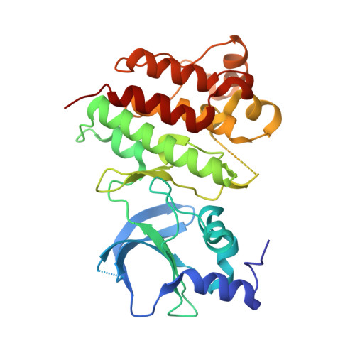
    8AN8, 8ANS

  • PubMed Abstract: 

    The c-MET receptor tyrosine kinase has received considerable attention as a cancer drug target yet there remains a need for inhibitors which are selective for c-MET and able to target emerging drug-resistant mutants. We report here the discovery, by screening a DNA-encoded chemical library, of a highly selective c-MET inhibitor which was shown by X-ray crystallography to bind to the kinase in an unprecedented manner. These results represent a novel mode of inhibiting c-MET with a small molecule and may provide a route to targeting drug-resistant forms of the kinase whilst avoiding potential toxicity issues associated with broad kinome inhibition.


  • Organizational Affiliation
    • Discovery Sciences, R&D, AstraZeneca, Cambridge, U.K. Electronic address: gavin.collie@astrazeneca.com.

Macromolecule Content 

  • Total Structure Weight: 34.01 kDa 
  • Atom Count: 2,227 
  • Modeled Residue Count: 277 
  • Deposited Residue Count: 296 
  • Unique protein chains: 1

Macromolecules

Find similar proteins by:|  3D Structure
Entity ID: 1
MoleculeChains  Sequence LengthOrganismDetailsImage
Hepatocyte growth factor receptor296Homo sapiensMutation(s): 1 
Gene Names: MET
EC: 2.7.10.1
UniProt & NIH Common Fund Data Resources
Find proteins for P08581 (Homo sapiens)
Explore P08581 
Go to UniProtKB:  P08581
PHAROS:  P08581
GTEx:  ENSG00000105976 
Entity Groups
Sequence Clusters30% Identity50% Identity70% Identity90% Identity95% Identity100% Identity
UniProt GroupP08581
Sequence Annotations
Expand
Reference Sequence

Small Molecules

Ligands 2 Unique
IDChains Name / Formula / InChI Key2D Diagram3D Interactions
MDI
(Subject of Investigation/LOI)

Query on MDI


Download:Ideal Coordinates CCD File
C [auth A]3-[bis(fluoranyl)methyl]-~{N}-methyl-~{N}-[(1~{R})-8-methyl-5-(3-methyl-1~{H}-indazol-6-yl)-1,2,3,4-tetrahydronaphthalen-1-yl]pyridine-2-carboxamide
C27 H26 F2 N4 O
ZGVMLURVURNUQI-HSZRJFAPSA-N
GOL

Query on GOL


Download:Ideal Coordinates CCD File
B [auth A]GLYCEROL
C3 H8 O3
PEDCQBHIVMGVHV-UHFFFAOYSA-N

Experimental Data & Validation

Experimental Data

  • Method: X-RAY DIFFRACTION
  • Resolution: 2.01 Å
  • R-Value Free:  0.277 (Depositor), 0.246 (DCC) 
  • R-Value Work:  0.229 (Depositor), 0.216 (DCC) 
  • R-Value Observed: 0.232 (Depositor) 
Space Group: P 21 21 21
Unit Cell:
Length ( Å )Angle ( ˚ )
a = 50.89α = 90
b = 52.83β = 90
c = 96.18γ = 90
Software Package:
Software NamePurpose
BUSTERrefinement
xia2data scaling
PDB_EXTRACTdata extraction
xia2data reduction
PHASERphasing

Structure Validation

View Full Validation Report


Ligand Structure Quality Assessment 


Entry History 

& Funding Information

Deposition Data

  • Released Date: 2022-08-31 
  • Deposition Author(s): Collie, G.W.

Funding OrganizationLocationGrant Number
Other privateUnited Kingdom--

Revision History  (Full details and data files)

  • Version 1.0: 2022-08-31
    Type: Initial release
  • Version 1.1: 2022-09-07
    Changes: Database references
  • Version 1.2: 2024-01-31
    Changes: Data collection, Refinement description