8AJ8

Structure of p110 gamma bound to the p84 regulatory subunit


Experimental Data Snapshot

  • Method: X-RAY DIFFRACTION
  • Resolution: 8.50 Å
  • R-Value Free: 0.340 
  • R-Value Work: 0.280 
  • R-Value Observed: 0.286 

wwPDB Validation   3D Report Full Report


This is version 1.1 of the entry. See complete history


Literature

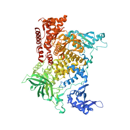
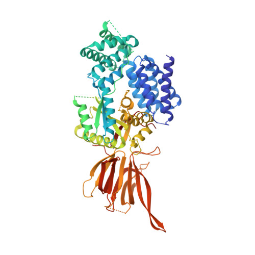
Molecular basis for differential activation of p101 and p84 complexes of PI3K gamma by Ras and GPCRs.

Rathinaswamy, M.K.Jenkins, M.L.Duewell, B.R.Zhang, X.Harris, N.J.Evans, J.T.Stariha, J.T.B.Dalwadi, U.Fleming, K.D.Ranga-Prasad, H.Yip, C.K.Williams, R.L.Hansen, S.D.Burke, J.E.

(2023) Cell Rep 42: 112172-112172

  • DOI: https://doi.org/10.1016/j.celrep.2023.112172
  • Primary Citation of Related Structures:  
    8AJ8

  • PubMed Abstract: 

    Class IB phosphoinositide 3-kinase (PI3Kγ) is activated in immune cells and can form two distinct complexes (p110γ-p84 and p110γ-p101), which are differentially activated by G protein-coupled receptors (GPCRs) and Ras. Using a combination of X-ray crystallography, hydrogen deuterium exchange mass spectrometry (HDX-MS), electron microscopy, molecular modeling, single-molecule imaging, and activity assays, we identify molecular differences between p110γ-p84 and p110γ-p101 that explain their differential membrane recruitment and activation by Ras and GPCRs. The p110γ-p84 complex is dynamic compared with p110γ-p101. While p110γ-p101 is robustly recruited by Gβγ subunits, p110γ-p84 is weakly recruited to membranes by Gβγ subunits alone and requires recruitment by Ras to allow for Gβγ activation. We mapped two distinct Gβγ interfaces on p101 and the p110γ helical domain, with differences in the C-terminal domain of p84 and p101 conferring sensitivity of p110γ-p101 to Gβγ activation. Overall, our work provides key insight into the molecular basis for how PI3Kγ complexes are activated.


  • Organizational Affiliation

    Department of Biochemistry and Microbiology, University of Victoria, Victoria, BC V8W 2Y2, Canada.


Macromolecules
Find similar proteins by:  (by identity cutoff)  |  3D Structure
Entity ID: 1
MoleculeChains Sequence LengthOrganismDetailsImage
Phosphatidylinositol 4,5-bisphosphate 3-kinase catalytic subunit gamma isoform
A, C, E, G
1,102Sus scrofaMutation(s): 0 
Gene Names: PIK3CG
EC: 2.7.1.137 (PDB Primary Data), 2.7.1.153 (PDB Primary Data), 2.7.1.154 (PDB Primary Data), 2.7.11.1 (PDB Primary Data)
UniProt
Find proteins for O02697 (Sus scrofa)
Explore O02697 
Go to UniProtKB:  O02697
Entity Groups  
Sequence Clusters30% Identity50% Identity70% Identity90% Identity95% Identity100% Identity
UniProt GroupO02697
Sequence Annotations
Expand
  • Reference Sequence
Find similar proteins by:  (by identity cutoff)  |  3D Structure
Entity ID: 2
MoleculeChains Sequence LengthOrganismDetailsImage
Phosphoinositide 3-kinase regulatory subunit 6
B, D, F, H
756Mus musculusMutation(s): 0 
Gene Names: Pik3r6
UniProt
Find proteins for Q3U6Q4 (Mus musculus)
Explore Q3U6Q4 
Go to UniProtKB:  Q3U6Q4
Entity Groups  
Sequence Clusters30% Identity50% Identity70% Identity90% Identity95% Identity100% Identity
UniProt GroupQ3U6Q4
Sequence Annotations
Expand
  • Reference Sequence
Experimental Data & Validation

Experimental Data

  • Method: X-RAY DIFFRACTION
  • Resolution: 8.50 Å
  • R-Value Free: 0.340 
  • R-Value Work: 0.280 
  • R-Value Observed: 0.286 
  • Space Group: C 1 2 1
Unit Cell:
Length ( Å )Angle ( ˚ )
a = 322.347α = 90
b = 166.598β = 114.024
c = 255.794γ = 90
Software Package:
Software NamePurpose
PHENIXrefinement
MOSFLMdata reduction
SCALAdata scaling
PHASERphasing

Structure Validation

View Full Validation Report


Entry History & Funding Information

Deposition Data


Funding OrganizationLocationGrant Number
Medical Research Council (MRC, United Kingdom)United KingdomMC_U105184308
Canadian Institutes of Health Research (CIHR)Canada168998

Revision History  (Full details and data files)

  • Version 1.0: 2023-03-22
    Type: Initial release
  • Version 1.1: 2024-02-07
    Changes: Data collection, Refinement description