8AHT

Crystal structure of Plasmodium falciparum Ca2+/Calmodulin in complex with melittin


Experimental Data Snapshot

  • Method: X-RAY DIFFRACTION
  • Resolution: 2.20 Å
  • R-Value Free: 0.266 
  • R-Value Work: 0.234 
  • R-Value Observed: 0.236 

wwPDB Validation   3D Report Full Report


This is version 1.2 of the entry. See complete history


Literature

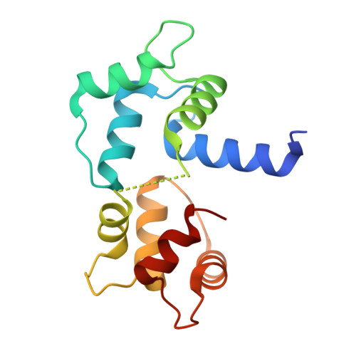
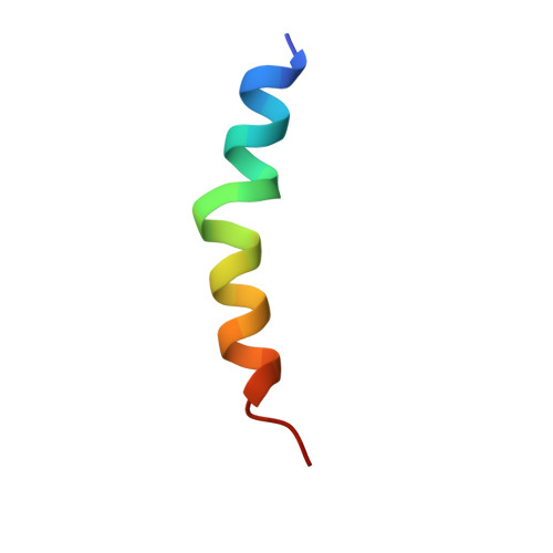
Structures of calmodulin-melittin complexes show multiple binding modes lacking classical anchoring interactions.

Durvanger, Z.Juhasz, T.Liliom, K.Harmat, V.

(2023) J Biol Chem 299: 104596-104596

  • DOI: https://doi.org/10.1016/j.jbc.2023.104596
  • Primary Citation of Related Structures:  
    8AHS, 8AHT

  • PubMed Abstract: 

    Calmodulin (CaM) is a Ca 2+ sensor protein found in all eukaryotic cells that regulates a large number of target proteins in a Ca 2+ concentration-dependent manner. As a transient-type hub protein, it recognizes linear motifs of its targets, though for the Ca 2+ -dependent binding, no consensus sequence was identified. Its complex with melittin, a major component of bee venom, is often used as a model system of protein-protein complexes. Yet, the structural aspects of the binding are not well understood, as only diverse, low-resolution data are available concerning the association. We present the crystal structure of melittin in complex with Ca 2+ -saturated CaMs from two, evolutionarily distant species, Homo sapiens and Plasmodium falciparum, representing three binding modes of the peptide. Results-augmented by molecular dynamics simulations-indicate that multiple binding modes can exist for CaM-melittin complexes, as an intrinsic characteristic of the binding. While the helical structure of melittin remains, swapping of its salt bridges and partial unfolding of its C-terminal segment can occur. In contrast to the classical way of target recognition by CaM, we found that different sets of residues can anchor at the hydrophobic pockets of CaM, which were considered as main recognition sites. Finally, the nanomolar binding affinity of the CaM-melittin complex is created by an ensemble of arrangements of similar stability-tight binding is achieved not by optimized specific interactions but by simultaneously satisfying less optimal interaction patterns in co-existing different conformers.


  • Organizational Affiliation

    Laboratory of Structural Chemistry and Biology, Institute of Chemistry, ELTE Eötvös Loránd University, Budapest, Hungary.


Macromolecules
Find similar proteins by:  (by identity cutoff)  |  3D Structure
Entity ID: 1
MoleculeChains Sequence LengthOrganismDetailsImage
Calmodulin
A, B, C, D
149Plasmodium falciparumMutation(s): 0 
Gene Names: PF14_0323
UniProt
Find proteins for P62203 (Plasmodium falciparum (isolate 3D7))
Explore P62203 
Go to UniProtKB:  P62203
Entity Groups  
Sequence Clusters30% Identity50% Identity70% Identity90% Identity95% Identity100% Identity
UniProt GroupP62203
Sequence Annotations
Expand
  • Reference Sequence
Find similar proteins by:  (by identity cutoff)  |  3D Structure
Entity ID: 2
MoleculeChains Sequence LengthOrganismDetailsImage
MelittinE [auth F],
F [auth G],
G [auth H],
H [auth I]
26Apis melliferaMutation(s): 0 
UniProt
Find proteins for P01501 (Apis mellifera)
Explore P01501 
Go to UniProtKB:  P01501
Entity Groups  
Sequence Clusters30% Identity50% Identity70% Identity90% Identity95% Identity100% Identity
UniProt GroupP01501
Sequence Annotations
Expand
  • Reference Sequence
Small Molecules
Ligands 1 Unique
IDChains Name / Formula / InChI Key2D Diagram3D Interactions
CA
Query on CA

Download Ideal Coordinates CCD File 
I [auth A]
J [auth A]
K [auth A]
L [auth A]
M [auth B]
I [auth A],
J [auth A],
K [auth A],
L [auth A],
M [auth B],
N [auth B],
O [auth B],
P [auth B],
Q [auth C],
R [auth C],
S [auth C],
T [auth C],
U [auth D],
V [auth D],
W [auth D],
X [auth D]
CALCIUM ION
Ca
BHPQYMZQTOCNFJ-UHFFFAOYSA-N
Experimental Data & Validation

Experimental Data

  • Method: X-RAY DIFFRACTION
  • Resolution: 2.20 Å
  • R-Value Free: 0.266 
  • R-Value Work: 0.234 
  • R-Value Observed: 0.236 
  • Space Group: P 1
Unit Cell:
Length ( Å )Angle ( ˚ )
a = 37.9α = 95.77
b = 60.68β = 89.99
c = 68.45γ = 97.05
Software Package:
Software NamePurpose
PHENIXrefinement
XDSdata reduction
XSCALEdata scaling
PHASERphasing

Structure Validation

View Full Validation Report


Entry History & Funding Information

Deposition Data


Funding OrganizationLocationGrant Number
Hungarian National Research, Development and Innovation OfficeHungary2018-1.2.1-NKP-2018-00005
European Regional Development FundEuropean UnionVEKOP-2.3.2-16-2017-00014, VEKOP-2.3.3-15-2017-00018
Hungarian National Research, Development and Innovation OfficeHungaryThematic Excellence Program Synth+

Revision History  (Full details and data files)

  • Version 1.0: 2023-03-22
    Type: Initial release
  • Version 1.1: 2023-04-26
    Changes: Database references
  • Version 1.2: 2024-02-07
    Changes: Data collection, Refinement description