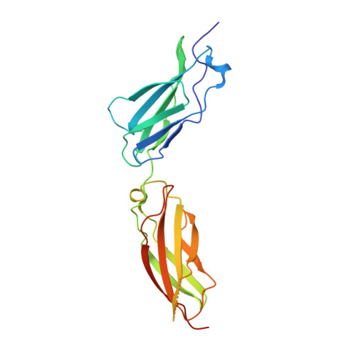
X-ray structure and function of fibronectin domains two and three of the neural cell adhesion molecule L1.
Guedez, G., Loers, G., Jeffries, C.M., Kozak, S., Meijers, R., Svergun, D.I., Schachner, M., Low, C.(2023) FASEB J 37: e22823-e22823
- PubMed: 36809668 Search on PubMed
- DOI: https://doi.org/10.1096/fj.202201511R
- Primary Citation Related Structures:
8AFO, 8AFP - PubMed Abstract:
The cell adhesion molecule L1 (L1CAM, L1 in short) plays crucial roles during neural development, regeneration after injury, synapse formation, synaptic plasticity and tumor cell migration. L1 belongs to the immunoglobulin superfamily and comprises in its extracellular part six immunoglobulin (Ig)-like domains and five fibronectin type III homologous repeats (FNs). The second Ig-like domain has been validated for self- (so-called homophilic) binding between cells. Antibodies against this domain inhibit neuronal migration in vitro and in vivo. The fibronectin type III homologous repeats FN2 and FN3 bind small molecule agonistic L1 mimetics and contribute to signal transduction. FN3 has a stretch of 25 amino acids that can be triggered with a monoclonal antibody, or the L1 mimetics, to enhance neurite outgrowth and neuronal cell migration in vitro and in vivo. To correlate the structural features of these FNs with function, we determined a high-resolution crystal structure of a FN2FN3 fragment, which is functionally active in cerebellar granule cells and binds several mimetics. The structure illustrates that both domains are connected by a short linker sequence allowing a flexible and largely independent organization of both domains. This becomes further evident by comparing the X-ray crystal structure with models derived from Small-Angle X-ray Scattering (SAXS) data for FN2FN3 in solution. Based on the X-ray crystal structure, we identified five glycosylation sites which we believe are crucial for folding and stability of these domains. Our study signifies an advance in the understanding of structure-functional relationships of L1.
- Centre for Structural Systems Biology (CSSB), Hamburg, Germany.
Organizational Affiliation: