8AEZ

X-ray structure of the deglycosylated receptor binding domain of Env glycoprotein of Simian Foamy virus


Experimental Data Snapshot

  • Method: X-RAY DIFFRACTION
  • Resolution: 2.57 Å
  • R-Value Free: 0.253 
  • R-Value Work: 0.213 
  • R-Value Observed: 0.215 

wwPDB Validation   3D Report Full Report


Ligand Structure Quality Assessment 


This is version 1.1 of the entry. See complete history


Literature

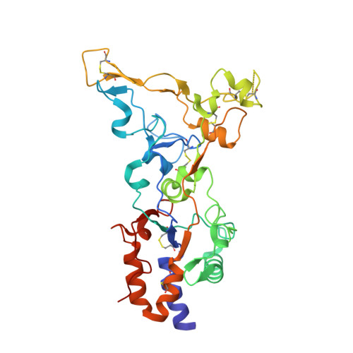
The crystal structure of a simian Foamy Virus receptor binding domain provides clues about entry into host cells.

Fernandez, I.Dynesen, L.T.Coquin, Y.Pederzoli, R.Brun, D.Haouz, A.Gessain, A.Rey, F.A.Buseyne, F.Backovic, M.

(2023) Nat Commun 14: 1262-1262

  • DOI: https://doi.org/10.1038/s41467-023-36923-0
  • Primary Citation of Related Structures:  
    8AEZ, 8AIC

  • PubMed Abstract: 

    The surface envelope glycoprotein (Env) of all retroviruses mediates virus binding to cells and fusion of the viral and cellular membranes. A structure-function relationship for the HIV Env that belongs to the Orthoretrovirus subfamily has been well established. Structural information is however largely missing for the Env of Foamy viruses (FVs), the second retroviral subfamily. In this work we present the X-ray structure of the receptor binding domain (RBD) of a simian FV Env at 2.57 Å resolution, revealing two subdomains and an unprecedented fold. We have generated a model for the organization of the RBDs within the trimeric Env, which indicates that the upper subdomains form a cage-like structure at the apex of the Env, and identified residues K342, R343, R359 and R369 in the lower subdomain as key players for the interaction of the RBD and viral particles with heparan sulfate.


  • Organizational Affiliation

    Institut Pasteur, Université Paris Cité, CNRS UMR3569, Unité de Virologie Structurale, 75015, Paris, France.


Macromolecules
Find similar proteins by:  (by identity cutoff)  |  3D Structure
Entity ID: 1
MoleculeChains Sequence LengthOrganismDetailsImage
Envelope glycoprotein gp130340Simian foamy virusMutation(s): 0 
Gene Names: env
UniProt
Find proteins for K7YEW5 (Simian foamy virus)
Explore K7YEW5 
Go to UniProtKB:  K7YEW5
Entity Groups  
Sequence Clusters30% Identity50% Identity70% Identity90% Identity95% Identity100% Identity
UniProt GroupK7YEW5
Sequence Annotations
Expand
  • Reference Sequence
Oligosaccharides

Help

Entity ID: 2
MoleculeChains Length2D Diagram Glycosylation3D Interactions
beta-D-mannopyranose-(1-4)-2-acetamido-2-deoxy-beta-D-glucopyranose-(1-4)-2-acetamido-2-deoxy-beta-D-glucopyranose
B
3N-Glycosylation
Glycosylation Resources
GlyTouCan:  G15407YE
GlyCosmos:  G15407YE
GlyGen:  G15407YE
Entity ID: 3
MoleculeChains Length2D Diagram Glycosylation3D Interactions
alpha-D-mannopyranose-(1-2)-alpha-D-mannopyranose-(1-2)-alpha-D-mannopyranose-(1-3)-[alpha-D-mannopyranose-(1-3)-alpha-D-mannopyranose-(1-6)]beta-D-mannopyranose-(1-4)-2-acetamido-2-deoxy-beta-D-glucopyranose-(1-4)-2-acetamido-2-deoxy-beta-D-glucopyranose
C
8N-Glycosylation
Glycosylation Resources
GlyTouCan:  G83582BK
GlyCosmos:  G83582BK
GlyGen:  G83582BK
Experimental Data & Validation

Experimental Data

  • Method: X-RAY DIFFRACTION
  • Resolution: 2.57 Å
  • R-Value Free: 0.253 
  • R-Value Work: 0.213 
  • R-Value Observed: 0.215 
  • Space Group: P 32 2 1
Unit Cell:
Length ( Å )Angle ( ˚ )
a = 99.451α = 90
b = 99.451β = 90
c = 120.609γ = 120
Software Package:
Software NamePurpose
BUSTERrefinement
XDSdata reduction
Aimlessdata scaling
AutoSolphasing

Structure Validation

View Full Validation Report


Ligand Structure Quality Assessment 


Entry History & Funding Information

Deposition Data

  • Released Date: 2023-02-22 
  • Deposition Author(s): Backovic, M.

Funding OrganizationLocationGrant Number
Laboratories of Excellence (LabEx)France--

Revision History  (Full details and data files)

  • Version 1.0: 2023-02-22
    Type: Initial release
  • Version 1.1: 2023-03-15
    Changes: Database references