8ADE | pdb_00008ade

Wild type ATTR amyloid fibril from senile systemic amyloidosis


Experimental Data Snapshot

  • Method: ELECTRON MICROSCOPY
  • Resolution: 2.78 Å
  • Aggregation State: HELICAL ARRAY 
  • Reconstruction Method: HELICAL 

wwPDB Validation 3D Report Full Report

Validation slider image for 8ADE

This is version 1.3 of the entry. See complete history

Literature

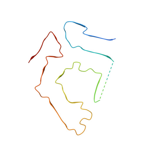
Cryo-EM structure of an ATTRwt amyloid fibril from systemic non-hereditary transthyretin amyloidosis.

Steinebrei, M.Gottwald, J.Baur, J.Rocken, C.Hegenbart, U.Schonland, S.Schmidt, M.

(2022) Nat Commun 13: 6398-6398

  • DOI: https://doi.org/10.1038/s41467-022-33591-4
  • Primary Citation Related Structures: 
    8ADE

  • PubMed Abstract: 

    Wild type transthyretin-derived amyloid (ATTRwt) is the major component of non-hereditary transthyretin amyloidosis. Its accumulation in the heart of elderly patients is life threatening. A variety of genetic variants of transthyretin can lead to hereditary transthyretin amyloidosis, which shows different clinical symptoms, like age of onset and pattern of organ involvement. However, in the case of non-hereditary transthyretin amyloidosis ATTRwt fibril deposits are located primarily in heart tissue. In this structural study we analyzed ATTRwt amyloid fibrils from the heart of a patient with non-hereditary transthyretin amyloidosis. We present a 2.78 Å reconstructed density map of these ATTRwt fibrils using cryo electron microscopy and compare it with previously published V30M variants of ATTR fibrils extracted from heart and eye of different patients. All structures show a remarkably similar spearhead like shape in their cross section, formed by the same N- and C-terminal fragments of transthyretin with some minor differences. This demonstrates common features for ATTR fibrils despite differences in mutations and patients.


  • Organizational Affiliation
    • Institute of Protein Biochemistry, Ulm University, Helmholtzstrasse 8/1, Ulm, D-89081, Germany.

Macromolecule Content 

  • Total Structure Weight: 96.44 kDa 
  • Atom Count: 5,026 
  • Modeled Residue Count: 644 
  • Deposited Residue Count: 889 
  • Unique protein chains: 1

Macromolecules

Find similar proteins by:|  3D Structure
Entity ID: 1
MoleculeChains  Sequence LengthOrganismDetailsImage
Transthyretin
A, B, C, D, E
A, B, C, D, E, F, G
127Homo sapiensMutation(s): 0 
UniProt & NIH Common Fund Data Resources
Find proteins for P02766 (Homo sapiens)
Explore P02766 
Go to UniProtKB:  P02766
PHAROS:  P02766
GTEx:  ENSG00000118271 
Entity Groups
Sequence Clusters30% Identity50% Identity70% Identity90% Identity95% Identity100% Identity
UniProt GroupP02766
Sequence Annotations
Expand
Reference Sequence

Experimental Data & Validation

Experimental Data

  • Method: ELECTRON MICROSCOPY
  • Resolution: 2.78 Å
  • Aggregation State: HELICAL ARRAY 
  • Reconstruction Method: HELICAL 

Structure Validation

View Full Validation Report


Entry History 

& Funding Information

Deposition Data


Funding OrganizationLocationGrant Number
German Research Foundation (DFG)GermanyFA 456/28

Revision History  (Full details and data files)

  • Version 1.0: 2022-11-23
    Type: Initial release
  • Version 1.1: 2022-11-30
    Changes: Structure summary
  • Version 1.2: 2023-03-15
    Changes: Advisory
  • Version 1.3: 2024-07-24
    Changes: Data collection