8AD2 | pdb_00008ad2

Tobacco lectin Nictaba in complex with triacetylchitotriose


Experimental Data Snapshot

  • Method: X-RAY DIFFRACTION
  • Resolution: 2.00 Å
  • R-Value Free: 
    0.226 (Depositor), 0.220 (DCC) 
  • R-Value Work: 
    0.196 (Depositor), 0.190 (DCC) 
  • R-Value Observed: 
    0.196 (Depositor) 

Starting Model: other
View more details

wwPDB Validation   3D Report Full Report


This is version 1.3 of the entry. See complete history


Literature

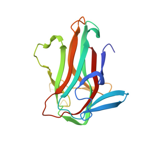
The crystal structure of Nictaba reveals its carbohydrate-binding properties and a new lectin dimerization mode.

Bloch, Y.Osterne, V.J.S.Savvides, S.N.Van Damme, E.J.M.

(2024) Glycobiology 

  • DOI: https://doi.org/10.1093/glycob/cwae087
  • Primary Citation of Related Structures:  
    8AD2, 8QMG

  • PubMed Abstract: 

    Nictaba is a (GlcNAc)n-binding, stress-inducible lectin from Nicotiana tabacum that serves as a representative for the Nictaba-related lectins, a group of proteins that play pivotal roles in plant defense mechanisms and stress response pathways. Despite extensive research into biological activities and physiological role(s) of the lectin, the three-dimensional structure of Nictaba remained largely unknown. Here, we report crystal structures for Nictaba in the apo form and bound to chitotriose. The structures reveal that the Nictaba protomer has a jelly-roll fold, similar to the cucumber lectin Cus17, but exhibit a unique and previously unseen mode of dimerization. The chitotriose binding mode, similar to Cus17, centers around the central GlcNAc residue, providing insights into the determinants of specificity of Nictaba towards carbohydrate structures. By integrating these structural insights with inputs from glycan arrays, molecular docking, and molecular dynamics simulations, we propose that Nictaba employs a single carbohydrate-recognition domain within each of the two subunits in the dimer to display pronounced specificity towards GlcNAc-containing carbohydrates. Furthermore, we identified amino acid residues involved in the extended binding site capable of accommodating structurally diverse high-mannose and complex N-glycans. Glycan array and in silico analyses revealed interactions centered around the conserved Man3GlcNAc2 core, explaining the broad recognition of N-glycan structures. Collectively, the structural and biochemical insights presented here fill a void into the atlas of lectin structure-function relationships and pave the way for future developments in plant stress biology and lectin-based applications.


  • Organizational Affiliation
    • Unit for Structural Biology, Department of Biochemistry and Microbiology, Ghent University, Technologiepark-Zwijnaarde 71, 9052 Ghent, Belgium.

Macromolecules
Find similar proteins by:  (by identity cutoff)  |  3D Structure
Entity ID: 1
MoleculeChains Sequence LengthOrganismDetailsImage
Nictaba
A, B, C, D, E
A, B, C, D, E, F, G, H
166Nicotiana tabacumMutation(s): 0 
Gene Names: NT1
UniProt
Find proteins for Q94EW1 (Nicotiana tabacum)
Explore Q94EW1 
Go to UniProtKB:  Q94EW1
Entity Groups  
Sequence Clusters30% Identity50% Identity70% Identity90% Identity95% Identity100% Identity
UniProt GroupQ94EW1
Sequence Annotations
Expand
  • Reference Sequence
Oligosaccharides

Help

Entity ID: 2
MoleculeChains Length2D Diagram Glycosylation3D Interactions
2-acetamido-2-deoxy-beta-D-glucopyranose-(1-4)-2-acetamido-2-deoxy-beta-D-glucopyranose-(1-4)-2-acetamido-2-deoxy-beta-D-glucopyranose
I, J, K, L
3N/A
Glycosylation Resources
GlyTouCan:  G47362BJ
GlyCosmos:  G47362BJ
GlyGen:  G47362BJ
Small Molecules
Ligands 2 Unique
IDChains Name / Formula / InChI Key2D Diagram3D Interactions
ZN
Query on ZN

Download Ideal Coordinates CCD File 
AA [auth E]
BA [auth E]
CA [auth E]
DA [auth F]
EA [auth F]
AA [auth E],
BA [auth E],
CA [auth E],
DA [auth F],
EA [auth F],
FA [auth F],
GA [auth F],
HA [auth G],
IA [auth G],
JA [auth G],
M [auth A],
O [auth B],
P [auth B],
Q [auth B],
R [auth B],
S [auth B],
T [auth C],
U [auth C],
V [auth C],
W [auth C],
X [auth D],
Y [auth D],
Z [auth D]
ZINC ION
Zn
PTFCDOFLOPIGGS-UHFFFAOYSA-N
CL
Query on CL

Download Ideal Coordinates CCD File 
N [auth A]CHLORIDE ION
Cl
VEXZGXHMUGYJMC-UHFFFAOYSA-M
Experimental Data & Validation

Experimental Data

  • Method: X-RAY DIFFRACTION
  • Resolution: 2.00 Å
  • R-Value Free:  0.226 (Depositor), 0.220 (DCC) 
  • R-Value Work:  0.196 (Depositor), 0.190 (DCC) 
  • R-Value Observed: 0.196 (Depositor) 
Space Group: P 1 21 1
Unit Cell:
Length ( Å )Angle ( ˚ )
a = 66.66α = 90
b = 146.159β = 95.874
c = 81.908γ = 90
Software Package:
Software NamePurpose
PHENIXrefinement
PHASERphasing
XDSdata reduction
XDSdata scaling

Structure Validation

View Full Validation Report


Entry History & Funding Information

Deposition Data


Funding OrganizationLocationGrant Number
Research Foundation - Flanders (FWO)Belgium12S0519N

Revision History  (Full details and data files)

  • Version 1.0: 2023-01-18
    Type: Initial release
  • Version 1.1: 2024-05-01
    Changes: Data collection, Refinement description
  • Version 1.2: 2024-05-22
    Changes: Database references
  • Version 1.3: 2024-11-20
    Changes: Database references, Structure summary