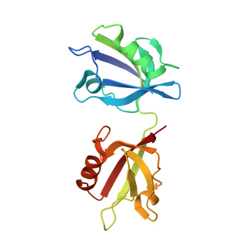
8AAO

Crystal structure of the PDZ tandem of syntenin in complex with compound 95


Experimental Data Snapshot

  • Method: X-RAY DIFFRACTION
  • Resolution: 2.47 Å
  • R-Value Free: 0.279 
  • R-Value Work: 0.203 
  • R-Value Observed: 0.206 

wwPDB Validation   3D Report Full Report


Ligand Structure Quality Assessment 


This is version 1.1 of the entry. See complete history


Literature

Discovery of a PDZ Domain Inhibitor Targeting the Syndecan/Syntenin Protein-Protein Interaction: A Semi-Automated "Hit Identification-to-Optimization" Approach.

Hoffer, L.Garcia, M.Leblanc, R.Feracci, M.Betzi, S.Ben Yaala, K.Daulat, A.M.Zimmermann, P.Roche, P.Barral, K.Morelli, X.

(2023) J Med Chem 66: 4633-4658

  • DOI: https://doi.org/10.1021/acs.jmedchem.2c01569
  • Primary Citation of Related Structures:  
    8AAI, 8AAK, 8AAO, 8AAP

  • PubMed Abstract: 

    The rapid identification of early hits by fragment-based approaches and subsequent hit-to-lead optimization represents a challenge for drug discovery. To address this challenge, we created a strategy called "DOTS" that combines molecular dynamic simulations, computer-based library design (chemoDOTS) with encoded medicinal chemistry reactions, constrained docking, and automated compound evaluation. To validate its utility, we applied our DOTS strategy to the challenging target syntenin, a PDZ domain containing protein and oncology target. Herein, we describe the creation of a "best-in-class" sub-micromolar small molecule inhibitor for the second PDZ domain of syntenin validated in cancer cell assays. Key to the success of our DOTS approach was the integration of protein conformational sampling during hit identification stage and the synthetic feasibility ranking of the designed compounds throughout the optimization process. This approach can be broadly applied to other protein targets with known 3D structures to rapidly identify and optimize compounds as chemical probes and therapeutic candidates.


  • Organizational Affiliation

    Centre de Recherche en Cancérologie de Marseille (CRCM), Aix-Marseille Université, Inserm 1068, CNRS 7258, Institut Paoli Calmettes, Marseille 13009, France.


Macromolecules
Find similar proteins by:  (by identity cutoff)  |  3D Structure
Entity ID: 1
MoleculeChains Sequence LengthOrganismDetailsImage
Syntenin-1
A, B
166Homo sapiensMutation(s): 0 
Gene Names: SDCBPMDA9SYCL
UniProt & NIH Common Fund Data Resources
Find proteins for O00560 (Homo sapiens)
Explore O00560 
Go to UniProtKB:  O00560
PHAROS:  O00560
GTEx:  ENSG00000137575 
Entity Groups  
Sequence Clusters30% Identity50% Identity70% Identity90% Identity95% Identity100% Identity
UniProt GroupO00560
Sequence Annotations
Expand
  • Reference Sequence
Small Molecules
Ligands 1 Unique
IDChains Name / Formula / InChI Key2D Diagram3D Interactions
LN2 (Subject of Investigation/LOI)
Query on LN2

Download Ideal Coordinates CCD File 
C [auth A],
D [auth B]
(2~{S})-2-[[(2~{S})-3-[4-(5-ethanoyl-2-fluoranyl-phenyl)phenyl]-2-(3-oxidanylidene-1~{H}-isoindol-2-yl)propanoyl]amino]propanoic acid
C28 H25 F N2 O5
TXGHAFIFHKHLOM-LMKMVOKYSA-N
Experimental Data & Validation

Experimental Data

  • Method: X-RAY DIFFRACTION
  • Resolution: 2.47 Å
  • R-Value Free: 0.279 
  • R-Value Work: 0.203 
  • R-Value Observed: 0.206 
  • Space Group: C 1 2 1
Unit Cell:
Length ( Å )Angle ( ˚ )
a = 102.206α = 90
b = 48.257β = 120.55
c = 74.942γ = 90
Software Package:
Software NamePurpose
SCALAdata scaling
BUSTERrefinement
PDB_EXTRACTdata extraction
autoPROCdata reduction
PHASERphasing

Structure Validation

View Full Validation Report


Ligand Structure Quality Assessment 


Entry History & Funding Information

Deposition Data


Funding OrganizationLocationGrant Number
Other governmentFrance--

Revision History  (Full details and data files)

  • Version 1.0: 2023-04-26
    Type: Initial release
  • Version 1.1: 2024-02-07
    Changes: Data collection, Refinement description