8AAN | pdb_00008aan

The nucleoprotein complex of Rep protein with iteron containing dsDNA and DUE ssDNA.


Experimental Data Snapshot

  • Method: X-RAY DIFFRACTION
  • Resolution: 2.19 Å
  • R-Value Free: 
    0.233 (Depositor), 0.225 (DCC) 
  • R-Value Work: 
    0.186 (Depositor) 
  • R-Value Observed: 
    0.189 (Depositor) 

Starting Model: experimental
View more details

wwPDB Validation 3D Report Full Report

Validation slider image for 8AAN

This is version 1.3 of the entry. See complete history

Literature

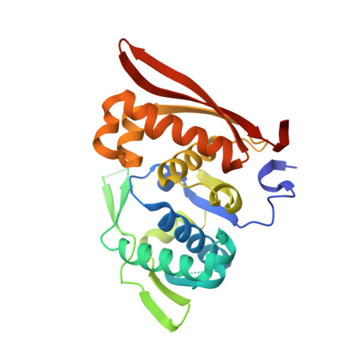
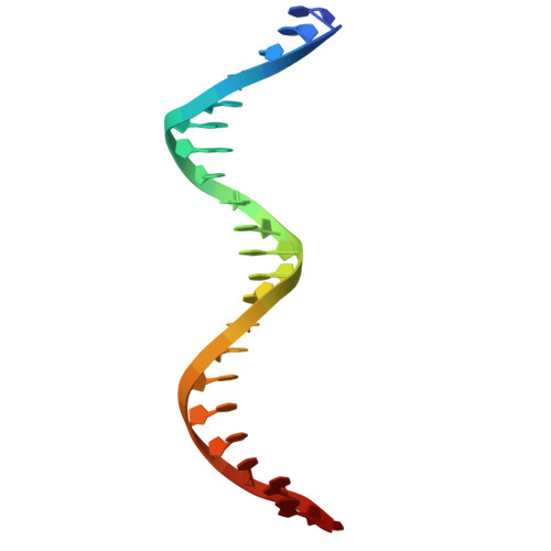
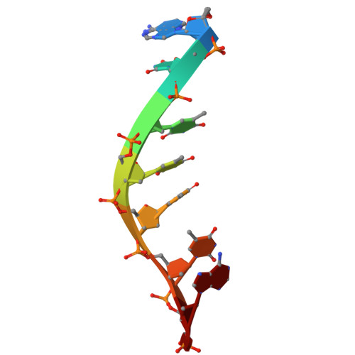
Rep protein accommodates together dsDNA and ssDNA which enables a loop-back mechanism to plasmid DNA replication initiation.

Wegrzyn, K.Oliwa, M.Nowacka, M.Zabrocka, E.Bury, K.Purzycki, P.Czaplewska, P.Pipka, J.Giraldo, R.Konieczny, I.

(2023) Nucleic Acids Res 51: 10551-10567

  • DOI: https://doi.org/10.1093/nar/gkad740
  • Primary Citation Related Structures: 
    8AAN, 8AC8

  • PubMed Abstract: 

    For DNA replication initiation in Bacteria, replication initiation proteins bind to double-stranded DNA (dsDNA) and interact with single-stranded DNA (ssDNA) at the replication origin. The structural-functional relationship of the nucleoprotein complex involving initiator proteins is still elusive and different models are proposed. In this work, based on crosslinking combined with mass spectrometry (MS), the analysis of mutant proteins and crystal structures, we defined amino acid residues essential for the interaction between plasmid Rep proteins, TrfA and RepE, and ssDNA. This interaction and Rep binding to dsDNA could not be provided in trans, and both are important for dsDNA melting at DNA unwinding element (DUE). We solved two crystal structures of RepE: one in a complex with ssDNA DUE, and another with both ssDNA DUE and dsDNA containing RepE-specific binding sites (iterons). The amino acid residues involved in interaction with ssDNA are located in the WH1 domain in stand β1, helices α1 and α2 and in the WH2 domain in loops preceding strands β1' and β2' and in these strands. It is on the opposite side compared to RepE dsDNA-recognition interface. Our data provide evidence for a loop-back mechanism through which the plasmid replication initiator molecule accommodates together dsDNA and ssDNA.


  • Organizational Affiliation
    • Intercollegiate Faculty of Biotechnology of University of Gdansk and Medical University of Gdansk, University of Gdansk, Abrahama 58, 80-307 Gdansk, Poland.

Macromolecule Content 

  • Total Structure Weight: 45.27 kDa 
  • Atom Count: 3,272 
  • Modeled Residue Count: 280 
  • Deposited Residue Count: 303 
  • Unique protein chains: 1
  • Unique nucleic acid chains: 3

Macromolecules


Find similar proteins by:|  3D Structure
Entity ID: 3
MoleculeChains  Sequence LengthOrganismDetailsImage
Replication initiation proteinC [auth A]251Escherichia coliMutation(s): 0 
Gene Names: repEErepECOK12F045
UniProt
Find proteins for P03856 (Escherichia coli (strain K12))
Explore P03856 
Go to UniProtKB:  P03856
Entity Groups
Sequence Clusters30% Identity50% Identity70% Identity90% Identity95% Identity100% Identity
UniProt GroupP03856
Sequence Annotations
Expand
Reference Sequence
Find similar nucleic acids by:  Sequence
Entity ID: 1
MoleculeChains LengthOrganismImage
DNA (5'-D(*CP*CP*TP*GP*TP*GP*AP*CP*AP*AP*AP*TP*TP*GP*CP*CP*CP*TP*CP*AP*GP*T)-3')A [auth B]22synthetic construct
Sequence Annotations
Expand
Reference Sequence
Find similar nucleic acids by:  Sequence
Entity ID: 2
MoleculeChains LengthOrganismImage
DNA (5'-D(*CP*TP*GP*AP*GP*GP*GP*CP*AP*AP*TP*TP*TP*GP*TP*CP*AP*CP*AP*GP*GP*T)-3')B [auth C]22synthetic construct
Sequence Annotations
Expand
Reference Sequence
Find similar nucleic acids by:  Sequence
Entity ID: 4
MoleculeChains LengthOrganismImage
DNA (5'-D(*AP*TP*TP*TP*TP*TP*AP*T)-3')8synthetic construct
Sequence Annotations
Expand
Reference Sequence

Small Molecules

Ligands 1 Unique
IDChains Name / Formula / InChI Key2D Diagram3D Interactions
MG
(Subject of Investigation/LOI)

Query on MG


Download:Ideal Coordinates CCD File
E [auth A]MAGNESIUM ION
Mg
JLVVSXFLKOJNIY-UHFFFAOYSA-N

Experimental Data & Validation

Experimental Data

  • Method: X-RAY DIFFRACTION
  • Resolution: 2.19 Å
  • R-Value Free:  0.233 (Depositor), 0.225 (DCC) 
  • R-Value Work:  0.186 (Depositor) 
  • R-Value Observed: 0.189 (Depositor) 
Space Group: C 1 2 1
Unit Cell:
Length ( Å )Angle ( ˚ )
a = 109.313α = 90
b = 81.047β = 123.52
c = 75.833γ = 90
Software Package:
Software NamePurpose
PHENIXrefinement
PDB_EXTRACTdata extraction
XDSdata reduction
XSCALEdata scaling
PHASERphasing

Structure Validation

View Full Validation Report


Entry History 

& Funding Information

Deposition Data


Funding OrganizationLocationGrant Number
Polish National Science CentrePoland2017/26/D/NZ1/00239

Revision History  (Full details and data files)

  • Version 1.0: 2023-09-27
    Type: Initial release
  • Version 1.1: 2023-10-04
    Changes: Structure summary
  • Version 1.2: 2023-11-08
    Changes: Database references
  • Version 1.3: 2026-03-04
    Changes: Refinement description, Structure summary