8AAM | pdb_00008aam

Crystal structure of MreB from Geobacillus stearothermophilus ATCC7953


Experimental Data Snapshot

  • Method: X-RAY DIFFRACTION
  • Resolution: 2.29 Å
  • R-Value Free: 
    0.274 (Depositor), 0.273 (DCC) 
  • R-Value Work: 
    0.202 (Depositor), 0.203 (DCC) 
  • R-Value Observed: 
    0.206 (Depositor) 

Starting Model: experimental
View more details

wwPDB Validation 3D Report Full Report

Validation slider image for 8AAM

Ligand Structure Quality Assessment 


This is version 1.1 of the entry. See complete history

Literature

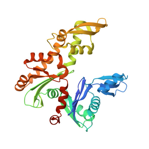
Polymerization cycle of an actin homolog MreB from a Gram-positive bacterium

Mao, W.Conilleau, C.Li de la Sierra-Gallay, I.Ah-Seng, Y.van Tilbeurgh, H.Renner, L.Nessler, S.Bertin, A.Chastanet, A.Carballido-Lopes, R.

To be published.

Macromolecule Content 

  • Total Structure Weight: 36.87 kDa 
  • Atom Count: 2,421 
  • Modeled Residue Count: 319 
  • Deposited Residue Count: 340 
  • Unique protein chains: 1

Macromolecules

Find similar proteins by:|  3D Structure
Entity ID: 1
MoleculeChains  Sequence LengthOrganismDetailsImage
Cell shape-determining protein MreBA [auth M]340Geobacillus stearothermophilusMutation(s): 0 
Gene Names: mreBB4109_2230B4114_2117GS458_2390
UniProt
Find proteins for A0A150MJ77 (Geobacillus stearothermophilus)
Explore A0A150MJ77 
Go to UniProtKB:  A0A150MJ77
Entity Groups
Sequence Clusters30% Identity50% Identity70% Identity90% Identity95% Identity100% Identity
UniProt GroupA0A150MJ77
Sequence Annotations
Expand
Reference Sequence

Small Molecules

Ligands 1 Unique
IDChains Name / Formula / InChI Key2D Diagram3D Interactions
ADP
(Subject of Investigation/LOI)

Query on ADP


Download:Ideal Coordinates CCD File
B [auth M]ADENOSINE-5'-DIPHOSPHATE
C10 H15 N5 O10 P2
XTWYTFMLZFPYCI-KQYNXXCUSA-N

Experimental Data & Validation

Experimental Data

  • Method: X-RAY DIFFRACTION
  • Resolution: 2.29 Å
  • R-Value Free:  0.274 (Depositor), 0.273 (DCC) 
  • R-Value Work:  0.202 (Depositor), 0.203 (DCC) 
  • R-Value Observed: 0.206 (Depositor) 
Space Group: P 1 21 1
Unit Cell:
Length ( Å )Angle ( ˚ )
a = 46.67α = 90
b = 61.71β = 112.17
c = 51.01γ = 90
Software Package:
Software NamePurpose
PHENIXrefinement
XDSdata reduction
XDSdata scaling
MOLREPphasing

Structure Validation

View Full Validation Report


Ligand Structure Quality Assessment 


Entry History 

& Funding Information

Deposition Data


Funding OrganizationLocationGrant Number
French Infrastructure for Integrated Structural Biology (FRISBI)FranceANR-10-INBS-05

Revision History  (Full details and data files)

  • Version 1.0: 2023-07-12
    Type: Initial release
  • Version 1.1: 2024-05-01
    Changes: Data collection, Refinement description