8A7T

beta-2-microglobulin D76N amyloid fibril form 2PFa


Experimental Data Snapshot

  • Method: ELECTRON MICROSCOPY
  • Resolution: 3.00 Å
  • Aggregation State: FILAMENT 
  • Reconstruction Method: HELICAL 

wwPDB Validation   3D Report Full Report


This is version 1.1 of the entry. See complete history


Literature

Disease-relevant beta 2 -microglobulin variants share a common amyloid fold.

Wilkinson, M.Gallardo, R.U.Martinez, R.M.Guthertz, N.So, M.Aubrey, L.D.Radford, S.E.Ranson, N.A.

(2023) Nat Commun 14: 1190-1190

  • DOI: https://doi.org/10.1038/s41467-023-36791-8
  • Primary Citation of Related Structures:  
    8A7O, 8A7P, 8A7Q, 8A7T

  • PubMed Abstract: 

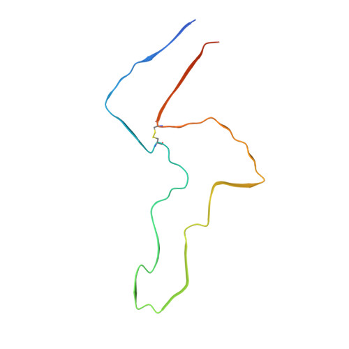
    β 2 -microglobulin (β 2 m) and its truncated variant ΔΝ6 are co-deposited in amyloid fibrils in the joints, causing the disorder dialysis-related amyloidosis (DRA). Point mutations of β 2 m result in diseases with distinct pathologies. β 2 m-D76N causes a rare systemic amyloidosis with protein deposited in the viscera in the absence of renal failure, whilst β 2 m-V27M is associated with renal failure, with amyloid deposits forming predominantly in the tongue. Here we use cryoEM to determine the structures of fibrils formed from these variants under identical conditions in vitro. We show that each fibril sample is polymorphic, with diversity arising from a 'lego-like' assembly of a common amyloid building block. These results suggest a 'many sequences, one amyloid fold' paradigm in contrast with the recently reported 'one sequence, many amyloid folds' behaviour of intrinsically disordered proteins such as tau and Aβ.


  • Organizational Affiliation

    Astbury Centre for Structural Molecular Biology, School of Molecular & Cellular Biology, Faculty of Biological Sciences, University of Leeds, Leeds, LS2 9JT, UK.


Macromolecules
Find similar proteins by:  (by identity cutoff)  |  3D Structure
Entity ID: 1
MoleculeChains Sequence LengthOrganismDetailsImage
Beta-2-microglobulin
A, B, C, D, E
A, B, C, D, E, F
99Homo sapiensMutation(s): 1 
Gene Names: B2MCDABP0092HDCMA22P
UniProt & NIH Common Fund Data Resources
Find proteins for P61769 (Homo sapiens)
Explore P61769 
Go to UniProtKB:  P61769
PHAROS:  P61769
GTEx:  ENSG00000166710 
Entity Groups  
Sequence Clusters30% Identity50% Identity70% Identity90% Identity95% Identity100% Identity
UniProt GroupP61769
Sequence Annotations
Expand
  • Reference Sequence
Experimental Data & Validation

Experimental Data

  • Method: ELECTRON MICROSCOPY
  • Resolution: 3.00 Å
  • Aggregation State: FILAMENT 
  • Reconstruction Method: HELICAL 
EM Software:
TaskSoftware PackageVersion
RECONSTRUCTIONRELION4.0
MODEL REFINEMENTPHENIX1.17

Structure Validation

View Full Validation Report


Entry History & Funding Information

Deposition Data


Funding OrganizationLocationGrant Number
Wellcome TrustUnited Kingdom--
Medical Research Council (MRC, United Kingdom)United Kingdom--
Royal SocietyUnited Kingdom--

Revision History  (Full details and data files)

  • Version 1.0: 2023-03-22
    Type: Initial release
  • Version 1.1: 2023-12-13
    Changes: Data collection, Refinement description