8A5R | pdb_00008a5r

Crystal structure of light-activated DNA-binding protein EL222 from Erythrobacter litoralis crystallized and measured in dark.


Experimental Data Snapshot

  • Method: X-RAY DIFFRACTION
  • Resolution: 1.85 Å
  • R-Value Free: 
    0.215 (Depositor), 0.180 (DCC) 
  • R-Value Work: 
    0.160 (Depositor), 0.170 (DCC) 
  • R-Value Observed: 
    0.161 (Depositor) 

Starting Model: experimental
View more details

wwPDB Validation   3D Report Full Report


Ligand Structure Quality Assessment 


This is version 1.2 of the entry. See complete history


Literature

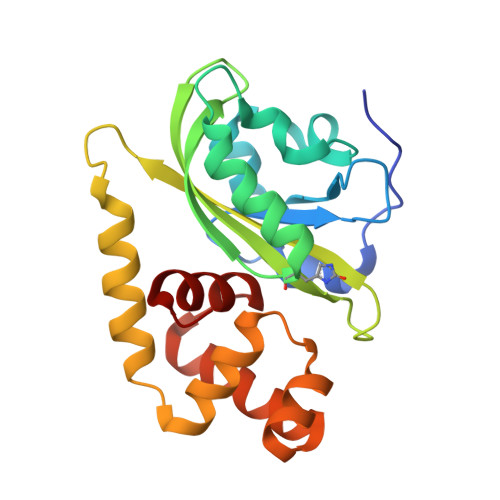
Light-dependent flavin redox and adduct states control the conformation and DNA-binding activity of the transcription factor EL222.

Chaudhari, A.S.Favier, A.Tehrani, Z.A.Koval, T.Andersson, I.Schneider, B.Dohnalek, J.Cerny, J.Brutscher, B.Fuertes, G.

(2025) Nucleic Acids Res 53

  • DOI: https://doi.org/10.1093/nar/gkaf215
  • Primary Citation of Related Structures:  
    8A5R, 8A5S

  • PubMed Abstract: 

    The activity of the light-oxygen-voltage/helix-turn-helix (LOV-HTH) photoreceptor EL222 is regulated through protein-protein and protein-DNA interactions, both triggered by photo-excitation of its flavin mononucleotide (FMN) cofactor. To gain molecular-level insight into the photocycle of EL222, we applied complementary methods: macromolecular X-ray crystallography (MX), nuclear magnetic resonance (NMR) spectroscopy, optical spectroscopies (infrared and UV-visible), molecular dynamics/metadynamics (MD/metaD) simulations, and protein engineering using noncanonical amino acids. Kinetic experiments provided evidence for two distinct EL222 conformations (lit1 and lit2) that become sequentially populated under illumination. These two lit states were assigned to covalently bound N5 protonated, and noncovalently bound hydroquinone forms of FMN, respectively. Only subtle structural differences were observed between the monomeric forms of all three EL222 species (dark, lit1, and lit2). While the dark state is largely monomeric, both lit states undergo monomer-dimer exchange. Furthermore, molecular modeling revealed differential dynamics and interdomain separation times arising from the three FMN states (oxidized, adduct, and reduced). Unexpectedly, all three EL222 species can associate with DNA, but only upon blue-light irradiation, a high population of stable complexes is obtained. Overall, we propose a model of EL222 activation where photoinduced changes in the FMN moiety shift the population equilibrium toward an open conformation that favors self-association and DNA-binding.


  • Organizational Affiliation
    • Laboratory of Biomolecular Recognition, Institute of Biotechnology of the Czech Academy of Sciences, Vestec 25250, Czech Republic.

Macromolecules
Find similar proteins by:  (by identity cutoff)  |  3D Structure
Entity ID: 1
MoleculeChains Sequence LengthOrganismDetailsImage
Light-activated DNA-binding protein EL222A [auth AAA]210Erythrobacter litoralis HTCC2594Mutation(s): 0 
Gene Names: ELI_04755
UniProt
Find proteins for Q2NB98 (Erythrobacter litoralis (strain HTCC2594))
Explore Q2NB98 
Go to UniProtKB:  Q2NB98
Entity Groups  
Sequence Clusters30% Identity50% Identity70% Identity90% Identity95% Identity100% Identity
UniProt GroupQ2NB98
Sequence Annotations
Expand
  • Reference Sequence
Small Molecules
Ligands 4 Unique
IDChains Name / Formula / InChI Key2D Diagram3D Interactions
FMN (Subject of Investigation/LOI)
Query on FMN

Download Ideal Coordinates CCD File 
B [auth AAA]FLAVIN MONONUCLEOTIDE
C17 H21 N4 O9 P
FVTCRASFADXXNN-SCRDCRAPSA-N
MES
Query on MES

Download Ideal Coordinates CCD File 
G [auth AAA]2-(N-MORPHOLINO)-ETHANESULFONIC ACID
C6 H13 N O4 S
SXGZJKUKBWWHRA-UHFFFAOYSA-N
CL
Query on CL

Download Ideal Coordinates CCD File 
C [auth AAA],
D [auth AAA]
CHLORIDE ION
Cl
VEXZGXHMUGYJMC-UHFFFAOYSA-M
MG
Query on MG

Download Ideal Coordinates CCD File 
E [auth AAA],
F [auth AAA]
MAGNESIUM ION
Mg
JLVVSXFLKOJNIY-UHFFFAOYSA-N
Modified Residues  1 Unique
IDChains TypeFormula2D DiagramParent
OHI
Query on OHI
A [auth AAA]L-PEPTIDE LINKINGC6 H7 N3 O3HIS
Experimental Data & Validation

Experimental Data

  • Method: X-RAY DIFFRACTION
  • Resolution: 1.85 Å
  • R-Value Free:  0.215 (Depositor), 0.180 (DCC) 
  • R-Value Work:  0.160 (Depositor), 0.170 (DCC) 
  • R-Value Observed: 0.161 (Depositor) 
Space Group: P 21 21 21
Unit Cell:
Length ( Å )Angle ( ˚ )
a = 43.81α = 90
b = 51.961β = 90
c = 81.063γ = 90
Software Package:
Software NamePurpose
PROTEUMdata collection
PROTEUMdata reduction
SADABSdata scaling
MOLREPphasing
Cootmodel building
REFMACrefinement

Structure Validation

View Full Validation Report


Ligand Structure Quality Assessment 


Entry History & Funding Information

Deposition Data


Funding OrganizationLocationGrant Number
Czech Academy of SciencesCzech Republic86652036
European Regional Development FundEuropean UnionCZ.02.1.01/0.0/0.0/15_003/0000447
Ministry of Education, Youth and Sports of the Czech RepublicCzech RepublicLM2018127

Revision History  (Full details and data files)

  • Version 1.0: 2023-07-05
    Type: Initial release
  • Version 1.1: 2024-02-07
    Changes: Data collection, Derived calculations, Refinement description
  • Version 1.2: 2025-04-09
    Changes: Database references, Structure summary